การประดิษฐ์อุปกรณ์ป้องกันรังสีจากยางธรรมชาติผสมบิสมัทปริมาณสูงสำหรับงานรังสีวินิจฉัย
คำสำคัญ:
การลดทอนปริมาณรังสี, บิสมัท, ยางธรรมชาติบทคัดย่อ
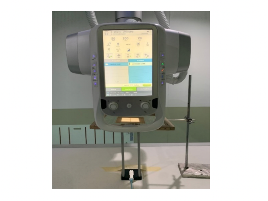
ปัจจุบันมีการนำรังสีเอกซ์มาใช้ประโยชน์ในทางการแพทย์ทั้งด้านการวินิจฉัยและการรักษา แต่อย่างไรก็ตามรังสีเอกซ์อาจจะก่อให้เกิดผลกระทบต่อร่างกาย เช่น คลื่นไส้ อาเจียนและการเพิ่มอัตราเสี่ยงการเกิดโรคมะเร็ง เป็นต้น ในการทำงานที่เกี่ยวข้องด้านรังสีจึงต้องมีการใช้อุปกรณ์ป้องกันรังสีอย่างเหมาะสมซึ่งโดยทั่วไปนิยมใช้ตะกั่วหรืออุปกรณ์ที่มีตะกั่วเป็นองค์ประกอบเพราะมีประสิทธิภาพในการลดทอนปริมาณรังสี ถึงอย่างไรก็ตามตะกั่วจัดเป็นสารที่อันตรายเนื่องจากในกระบวนการผลิตอาจจะปล่อยของเสียที่เป็นอันตรายแก่สิ่งแวดล้อมและนำไปสู่การเกิดปัญหาสุขภาพได้ งานวิจัยนี้มีวัตถุประสงค์เพื่อประดิษฐ์แผ่นป้องกันรังสีที่มีส่วนผสมของยางธรรมชาติและบิสมัทปริมาณสูงทดแทนการใช้ตะกั่วเป็นส่วนผสม และเปรียบเทียบประสิทธิภาพในการลดทอนรังสีของแผ่นป้องกันรังสีที่มีส่วนผสมของยางแท่งและบิสมัทปริมาณต่าง ๆ กับอุปกรณ์ป้องกันรังสีที่มีสมมูลตะกั่ว 0.5 มิลลิเมตร โดยทำการทดลองตัวอย่างแผ่นยางทีเตรียมจากสูตรยางธรรมชาติผสมบิสมัททั้งหมด 6 สูตร คือ 0, 100, 200, 300, 400 และ 500 phr (หน่วยการผสมยางโดยคิดสัดส่วนปริมาณสารต่าง ๆ เมื่อเทียบกับยาง 100 ส่วน โดยน้ำหนัก) โดยที่ตัวอย่างแผ่นยางมีความหนาตั้งแต่ 1 ถึง 5 มิลลิเมตร ทำการทดสอบประสิทธิภาพในการลดทอนปริมาณรังสีโดยใช้ค่าเอกซโพเชอร์ที่ความต่างศักย์ในช่วง 50 ถึง 120 เควีพี และค่ากระแสหลอด-เวลา 20 เอ็มเอเอส จากผลการทดลองพบว่าร้อยละการลดทอนปริมาณรังสีมีค่าเพิ่มขึ้นตามปริมาณของบิสมัทที่เพิ่มขึ้น เมื่อเปรียบเทียบกับอุปกรณ์ป้องกันรังสีที่มีสมมูลตะกั่ว 0.5 มิลลิเมตร กับแผ่นป้องกันรังสีที่ประดิษฐ์ขึ้นพบว่าแผ่นยางสูตรที่ผสมตะกั่ว 300 phr ที่มีความหนา 3 มิลลิเมตร มีค่าร้อยละการลดทอนปริมาณรังสีเท่ากับ 97.33 ที่ 100 เควีพี ซึ่งใกล้เคียงกับกับอุปกรณ์ป้องกันรังสีที่มีความหนาสมมูลตะกั่ว 0.5 มิลลิเมตร และเมื่อเพิ่มปริมาณบิสมัทเป็น 400 และ 500 phr ประสิทธิภาพการลดทอนปริมาณรังสีได้ดีกว่าอุปกรณ์เปรียบเทียบที่มีความหนา 3 มิลลิเมตร
Downloads
เอกสารอ้างอิง
Mettler FA. Medical effects and risks of exposure to ionising radiation. J Radiol Prot 2012; 32(1): 9-13.
Wagner LK, Eifel PJ, Geise RA. Potential Biological Effects Following High X-ray Dose Interventional Procedures. J Vasc Interv Radiol 1994; 5(1): 71-84.
ราชกิจจานุเบกษา. พระราชบัญญัติพลังงานนิวเคลียร์ พ.ศ.๒๕๕๙. [อินเตอร์เน็ต] [เข้าถึงเมื่อ ๑๒ ธ.ค. ๒๕๕๙] เข้าถึงได้จาก; http://www.oap.go.th/images/documents/about-us/regulations/nuclear-energy-act-2559.PDF.
Sudjai W. Inlight optically stimulated luminescence for occupational monitoring service in Thailand. Bull Dept Med Sci 2012; 54(2): 110-116.
Agyingi EO, Mobit PN, Sandison GA. Energy response of an aluminium oxide detector in kilovoltage and megavoltage photon beams: an EGSnrc Monte Carlo simulation study. Radiat Prot Dosimetry 2006; 118(1): 28-31.
McKeever SWS, Akselrod MS. Radiation dosimetry using pulsed optically stimulated luminescence of Al2O3:C. Radiat Prot Dosimetry 1999; 84(1-4): 317-320.
Bauhs JA, Vrieze TJ, Primak AN, Bruesewitz MR, McCollough CH. CT dosimetry: comparison of measurement techniques and devices. Radiographics 2008; 28(1): 245-253.
Landauer Inc. Dosimeters: InLight® Systems Dosimeters. [Internet]. 2005. [cited 2017 May 20]. Available from: https://www.nagase-landauer.co.jp/english/inlight/pdf/Dosimeters/inlightdosimeters.pdf
ยุทธนา บางม่วง, วราภรณ์ สุดใจ. การสำรวจปริมาณรังสีพื้นหลังของประเทศโดยใช้แผ่นวัดรังสีชนิดโอเอสแอล. วารสารกรมวิทยาศาสตร์การแพทย์. ๒๕๕๖;๕๕:๓๑-๓๘.

ดาวน์โหลด
เผยแพร่แล้ว
รูปแบบการอ้างอิง
ฉบับ
ประเภทบทความ
สัญญาอนุญาต
บทความที่ได้รับการตีพิมพ์เป็นลิขสิทธิ์ของสมาคมรังสีเทคนิคแห่งประเทศไทย (The Thai Society of Radiological Technologists)
ข้อความที่ปรากฏในบทความแต่ละเรื่องในวารสารวิชาการเล่มนี้เป็นความคิดเห็นส่วนตัวของผู้เขียนแต่ละท่านไม่เกี่ยวข้องกับสมาคมรังสีเทคนิคแห่งประเทศไทยและบุคคลากรท่านอื่น ๆในสมาคม ฯ แต่อย่างใด ความรับผิดชอบองค์ประกอบทั้งหมดของบทความแต่ละเรื่องเป็นของผู้เขียนแต่ละท่าน หากมีความผิดพลาดใดๆ ผู้เขียนแต่ละท่านจะรับผิดชอบบทความของตนเองแต่ผู้เดียว


