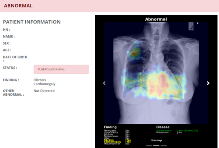
Evaluation of pulmonary tuberculosis diagnosis in chest radiographs using the AXIR-CX artificial intelligence program from confirmation results based on the Xpert MTB/RIF Ultra method in recipients with negative AFB sputum results
Keywords:
Chest digital radiograph, AXIR-CX Artificial Intelligence Program, Xpert MTB/RIF Ultra Method, Pulmonary tuberculosisAbstract
Introduction: Tuberculosis is a problem for the public health system both globally and in Thailand. When screening for pulmonary tuberculosis using radiographic images, a shortage of radiologists to interpret the images was found. Therefore, the AXIR-CX artificial intelligence program was introduced for image interpretation. This study aims to evaluate the performance of the AXIR-CX artificial intelligence program in screening for pulmonary tuberculosis using digital chest radiographs, with the Xpert MTB/RIF Ultra method as the reference standard. Methods: A retrospective descriptive design was used, with digital chest radiographs randomly selected from samples at six primary hospitals in Chanthaburi province using the AeroDR 2 1417S DR detector. Data were collected from October 1, 2019, to September 30, 2023. The data were analyzed using diagnostic test statistics, ROC curve analysis, and Kappa statistics for agreement measurement. Results: The AXIR-CX artificial intelligence program analyzed 459 chest X-ray images and found that the sensitivity and specificity were 80.6% (95% CI: 69.5-88.9) and 59.2% (95% CI: 54.1-64.1), respectively. The accuracy was 62.5% (95% CI: 58.1-66.9). The positive likelihood ratio was 1.97 (95%CI: 1.67 to 2.33), and the negative likelihood ratio was 0.33 (95%CI: 0.20 to 0.53). The ROC curve analysis yielded a cutoff point of 0.5, with the area under the ROC curve being 69.9% (95% CI: 64.6-75.1). When setting the significance level at 0.05, there was no statistically significant difference in the area under the ROC curve between males and females (p-value = 0.12), and there was no statistically significant difference in the area under the ROC curve between the age groups of 15-54 years and those aged 55 years and older (p-value = 0.29). The kappa coefficient between the AXIR-CX artificial intelligence program and the Xpert MTB/RIF Ultra method was 0.22 (95% CI: 0.15-0.29). Conclusion: The AXIR-CX artificial intelligence program can be used with caution for screening pulmonary tuberculosis in primary hospitals, as the decision criteria for estimating the area under the ROC curve is relatively low, and the level of agreement strength is fair.
Downloads
References
World Health Organization. Annual report of tuberculosis. Annual global TB report of WHO [Internet]. 2022 [cited 2023 Sep 22];8(1):1-68.
Provisional tuberculosis (TB) notifications [Internet]. Geneva: World Health Organization; 2023 [cited 2023 Sep 22].
กองวัณโรค กรมควบคุมโรค. National Tuberculosis Control Programme Guideline, Thailand 2021. Bangkok: Division of Tuberculosis; 2021.
World Health Organization. Systematic screening for active tuberculosis: an operational guide. Geneva: World Health Organization; 2015.
World Health Organization. WHO meeting report of a technical expert consultation: non-inferiority analysis of Xpert MTB/RIF Ultra compared to Xpert MTB/RIF. Geneva: World Health Organization; 2017. Report No.: WHO/HTM/TB/2017.04. Licence: CC BY-NC-SA 3.0 IGO.
World Health Organization. WHO consolidated guidelines on tuberculosis. Module 3: Diagnosis. In: Rapid diagnostics for tuberculosis detection. Geneva: World Health Organization; 2020.
ระบบสารสนเทศภูมิศาสตร์ทรัพยากรสุขภาพ [Internet]. [cited 2023 Sep 22].
Yoo SH, Geng H, Chiu TL, Yu SK, Cho DC, Heo J, et al. Study on the TB and non-TB diagnosis using two-step deep learning-based binary classifier. J Instrum. 2020 Oct 1;15(10).
Codlin AJ, Dao TP, Vo LNQ, Forse RJ, Van Truong V, Dang HM, et al. Independent evaluation of 12 artificial intelligence solutions for the detection of tuberculosis. Sci Rep. 2021 Dec 1;11(1).
Hajian-Tilaki K. Sample size estimation in diagnostic test studies of biomedical informatics. J Biomed Inform. 2014;48:193–204.
Zhan Y, Wang Y, Zhang W, Ying B, Wang C. Diagnostic accuracy of the artificial intelligence methods in medical imaging for pulmonary tuberculosis: A systematic review and meta-analysis. J Clin Med. 2023;12(1):303.
สำนักวัณโรค กรมควบคุมโรค กระทรวงสาธารณสุข. การสำรวจความชุกของวัณโรคระดับชาติในประเทศไทย ปี พ.ศ. 2555-2556. 2560.
Chu K. An introduction to sensitivity, specificity, predictive values and likelihood ratios. Emerg Med. 1999;11(3):175-181.
พงษ์เดช สารการ. หลักการทางชีวสถิติและแนวทางการวิเคราะห์ข้อมูลด้วยโปรแกรม STATA 15. ขอนแก่น: สาขาวิชาวิทยาการระบาดและชีวสถิติ คณะสาธารณสุขศาสตร์ มหาวิทยาลัยขอนแก่น; 2566.
Landis JR, Koch GG. The measurement of observer agreement for categorical data. Biometrics. 1997;33(1):159-174.
van’t Hoog A, Viney K, Biermann O, Yang B, Leeflang MMG, Langendam MW. Symptom- and chest-radiography screening for active pulmonary tuberculosis in HIV-negative adults and adults with unknown HIV status. Cochrane Database of Systematic Reviews. 2022;3.

Downloads
Published
How to Cite
Issue
Section
License
Copyright (c) 2024 The Thai Society of Radiological Technologists

This work is licensed under a Creative Commons Attribution-NonCommercial-NoDerivatives 4.0 International License.
บทความที่ได้รับการตีพิมพ์เป็นลิขสิทธิ์ของสมาคมรังสีเทคนิคแห่งประเทศไทย (The Thai Society of Radiological Technologists)
ข้อความที่ปรากฏในบทความแต่ละเรื่องในวารสารวิชาการเล่มนี้เป็นความคิดเห็นส่วนตัวของผู้เขียนแต่ละท่านไม่เกี่ยวข้องกับสมาคมรังสีเทคนิคแห่งประเทศไทยและบุคคลากรท่านอื่น ๆในสมาคม ฯ แต่อย่างใด ความรับผิดชอบองค์ประกอบทั้งหมดของบทความแต่ละเรื่องเป็นของผู้เขียนแต่ละท่าน หากมีความผิดพลาดใดๆ ผู้เขียนแต่ละท่านจะรับผิดชอบบทความของตนเองแต่ผู้เดียว


