
Evaluation of CT calibration curve impact on proton range accuracy in treatment planning
Keywords:
CT number, Proton therapy, Calibration curve, Proton Stopping Power, Treatment Planning SystemAbstract
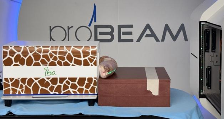
Introduction: In proton therapy, the accuracy of dose calculation and proton range determination is critically dependent on the conversion of CT number to proton stopping power ratio (SPR). Any inaccuracy in the CT calibration curve can lead to proton range uncertainties and potential deviations in dose distribution within the target and surrounding organs at risk. Therefore, verification of the accuracy of the CT calibration curve used in treatment planning is essential to ensure precise dose delivery. Objective: To evaluate the accuracy and reliability of the CT calibration curve currently implemented in the proton therapy treatment planning system at the Department of Radiology, King Chulalongkorn Memorial Hospital, The Thai Red Cross Society. Methods: A calibration curve was generated by correlating CT numbers with the proton stopping powers of Gammex tissue-equivalent materials. The resulting curve was applied in the treatment planning system to calculate proton ranges, which were then compared with measurements obtained using a Giraffe dosimeter on a Varian ProBeam system. Both phantom and real tissue samples were evaluated for comparison. Results: No statistically significant difference was found between calculated and measured proton ranges (P = 0.21). The mean differences of R80 and R90 in the phantom and real tissue were 1.53 ± 3.85 mm and 0.51 ± 3.08 mm, and 1.54 ± 3.82 mm and 0.83 ± 2.43 mm, respectively. Six of the Gammex materials met the AAPM TG-185 criteria, while the remaining materials showed consistent results with previous studies using the same CT and proton systems. Conclusion: The current CT calibration curve used in the proton therapy planning system at King Chulalongkorn Memorial Hospital demonstrates acceptable accuracy and reliability for clinical dose calculation and treatment planning applications.
Downloads
References
Farr JB, Moyers MF, Allgower CE, Bues M, Hsi WC, Jin H, et al. Clinical commissioning of intensity-modulated proton therapy systems: report of AAPM Task Group 185. Med Phys. 2021;48(1):e1–e30. doi:10.1002/mp.14546.
Schneider U, Pedroni E, Lomax A. The calibration of CT Hounsfield units for radiotherapy treatment planning. Phys Med Biol. 1996;41(1):111–124.
Yang M, Zhu XR, Park PC, Titt U, Mohan R, Virshup G, et al. Comprehensive analysis of proton range uncertainties related to patient stopping-power-ratio estimation using the stoichiometric calibration. Med Phys. 2010;37(8):3674–3684.
Moyers MF, Miller DW, Bush DA, Slater JD. Methodologies and tools for proton beam verification and validation. Radiat Oncol. 2016;11:118.
Bethe HA. Zur Theorie des Durchgangs schneller Korpuskularstrahlen durch Materie. Ann Phys. 1930;397(5):325–400. doi:10.1002/andp.19303970303.
International Commission on Radiation Units and Measurements. ICRU Report 90: Key Data for Ionizing-Radiation Dosimetry: Measurement Standards and Applications. Bethesda (MD): ICRU; 2016.
Monkongsubsin W, Israngkul Na Ayuthaya I, Sanghangthum T, Keawsamur M. Range comparison of Monte Carlo and pencil beam algorithms in treatment planning system for proton therapy. In: Proceedings of the Thai Medical Physics Society Conference; 2024; Bangkok, Thailand. Bangkok: TMPS; 2024. p. 1–6.
Paganetti H. Range uncertainties in proton therapy and the role of Monte Carlo simulations. Phys Med Biol. 2012;57(11):R99–R117.
Dennis ML, Radovich E, Wong KLM, Owolabi O, Cavallaro FL, Mbizvo MT, et al. Pathways to increased coverage: an analysis of time trends in contraceptive need and use among adolescents and young women in Kenya, Rwanda, Tanzania, and Uganda. Reprod Health. 2017;14(1):130. doi:10.1186/s12978-017-0393-3.
Chirdchid T, Ruangchan S, Sanghangthum T. Dosimetric comparison between single-energy computed tomography and dual-energy computed tomography relative to stopping power estimation in proton therapy. J Med Phys. 2023;48(3):292–297. doi:10.4103/jmp.jmp_67_23.
Downloads
Published
How to Cite
Issue
Section
License
Copyright (c) 2025 The Thai Society of Radiological Technologists

This work is licensed under a Creative Commons Attribution-NonCommercial-NoDerivatives 4.0 International License.
บทความที่ได้รับการตีพิมพ์เป็นลิขสิทธิ์ของสมาคมรังสีเทคนิคแห่งประเทศไทย (The Thai Society of Radiological Technologists)
ข้อความที่ปรากฏในบทความแต่ละเรื่องในวารสารวิชาการเล่มนี้เป็นความคิดเห็นส่วนตัวของผู้เขียนแต่ละท่านไม่เกี่ยวข้องกับสมาคมรังสีเทคนิคแห่งประเทศไทยและบุคคลากรท่านอื่น ๆในสมาคม ฯ แต่อย่างใด ความรับผิดชอบองค์ประกอบทั้งหมดของบทความแต่ละเรื่องเป็นของผู้เขียนแต่ละท่าน หากมีความผิดพลาดใดๆ ผู้เขียนแต่ละท่านจะรับผิดชอบบทความของตนเองแต่ผู้เดียว


