
Establishment of typical values for computed tomography simulation at Sawanpracharak Hospital
Keywords:
Computed tomography simulator, Volume CT dose index (CTDIvol), Dose length product (DLP), Diagnostic reference levels (DRLs)Abstract
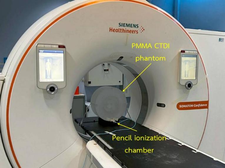
Background: Diagnostic reference levels (DRLs) serve as essential tools for monitoring and optimizing radiation exposure in computed tomography (CT). At the institutional level, typical values defined as the median of the dose distribution can be used as benchmarks for establishing and reviewing DRLs. Objective: To establish institutional typical values for a CT simulator at Sawanpracharak Hospital. Methods: A total of 292 patients who underwent CT simulation using a Siemens SOMATOM Confidence RT Pro between January and December 2024 were included. Patient characteristics, scan parameters, and dose indices—volume CT dose index (CTDIvol) and dose length product (DLP)—were collected across five anatomical regions: brain, head–neck, chest, abdomen, and pelvis. Typical values were defined as the median of the dose distributions, and correlations between DLP and patient factors as well as scan parameters were analyzed. Results: Dose levels varied by region, with the highest typical values of CTDIvol and DLP observed in the brain (68.83 mGy, 2173.05 mGy·cm) and the lowest in the head–neck region (7.24 mGy, 325.00 mGy·cm), while the chest, abdomen, and pelvis showed comparable levels. Strong positive correlations were found between DLP and both mAs and scan length across all regions (p<0.001). Age showed no significant correlation, whereas BMI correlated only in the chest, abdomen, and pelvis. Pitch demonstrated significant associations in all regions except the brain. Conclusion: The derived typical values can serve as baseline data for the future development of DRLs for CT simulation, thereby enhancing radiation safety in radiotherapy treatment planning.
Downloads
References
ศุภวิทู สุขเพ็ง. เทคนิคการถ่ายภาพเอกซเรย์ซีทีด้วยปริมาณรังสีที่เหมาะสม. พิษณุโลก: สำนักพิมพ์มหาวิทยาลัยนเรศวร; 2565.
Sodkokkruad P, Asavaphatiboon S, Thanabodeebonsiri J, Tangboonduangjit P. Comparison of computed tomography dose index measuring by two detector types of computed tomography simulator. Thai J Rad Tech. 2018;43(1):64-8.
International Commission on Radiological Protection. The 2007 recommendations of the International Commission on Radiological Protection (ICRP Publication 103). Ann ICRP. 2007;37(2–4):1–332.
International Commission on Radiological Protection. Diagnostic reference levels in medical imaging (ICRP Publication 135). Ann ICRP. 2017;46(1):1–144.
กรมวิทยาศาสตร์การแพทย์ กระทรวงสาธารณสุข. ค่าปริมาณรังสีอ้างอิงในการถ่ายภาพรังสีวินิจฉัยทางการแพทย์ของประเทศไทย พ.ศ. 2564. นนทบุรี: บริษัท บียอนด์ พับลิสซิ่ง จำกัด; 2564.
Donmoon T, Chusin T. Establishment of typical dose levels for computed tomography of the pelvis region in radiotherapy simulation procedures. Journal of Thai Association of Radiation Oncology 2023;29(1):23-33.
Zalokar N, Žager Marciuš V, Mekiš N. Establishment of national diagnostic reference levels for radiotherapy computed tomography simulation procedures in Slovenia. Eur J Radiol. 2020;127:108979.
International Atomic Energy Agency. Quality assurance for computed tomography: diagnostic and therapy applications. Human Health Series No. 19. Vienna: IAEA; 2012.
Nayak SS, Yadav S, Pradhan A. Effect of body mass index on effective dose in multidetector computed tomography abdomen using automatic exposure control. Ethiop J Health Sci. 2024;34(6):494–500.
Pimsorn P. Factors affecting size-specific dose estimates in addition to scanning parameters for chest-abdomen-pelvis computed tomography using automatic tube current modulation at Chiangrai prachanukroh hospital. Thai J Rad Tech. 2022;47(1):43-54.
Donmoon T, Chusin T. Establishment of local diagnostic reference levels for commonly performed computed tomography examinations in Thai cancer hospitals. Thai J Rad Tech. 2021;46(1):35-42.
Ruenjit S, Siricharoen P, Khamwan K. Automated size-specific dose estimates framework in thoracic CT using a convolutional neural network based on a U-Net model. J Appl Clin Med Phys. 2024;25(3):e14283.
Downloads
Published
How to Cite
Issue
Section
License
Copyright (c) 2025 The Thai Society of Radiological Technologists

This work is licensed under a Creative Commons Attribution-NonCommercial-NoDerivatives 4.0 International License.
บทความที่ได้รับการตีพิมพ์เป็นลิขสิทธิ์ของสมาคมรังสีเทคนิคแห่งประเทศไทย (The Thai Society of Radiological Technologists)
ข้อความที่ปรากฏในบทความแต่ละเรื่องในวารสารวิชาการเล่มนี้เป็นความคิดเห็นส่วนตัวของผู้เขียนแต่ละท่านไม่เกี่ยวข้องกับสมาคมรังสีเทคนิคแห่งประเทศไทยและบุคคลากรท่านอื่น ๆในสมาคม ฯ แต่อย่างใด ความรับผิดชอบองค์ประกอบทั้งหมดของบทความแต่ละเรื่องเป็นของผู้เขียนแต่ละท่าน หากมีความผิดพลาดใดๆ ผู้เขียนแต่ละท่านจะรับผิดชอบบทความของตนเองแต่ผู้เดียว


