
Livedo reticularis-an unusual skin manifestation of disseminated strongyloidiasis: a case report with literature review
Keywords:
Disseminated strongyloidiasis, thumbprint purpura, livedo reticularisAbstract
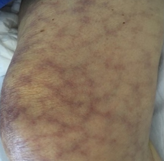
A 71-year-old woman, with active autoimmune hepatitis, was treated with immunosuppressive drugs and presented with a1-month history of fever and diarrhea, dyspnea, and sudden eruptions of purpuric macules on the abdomen typical for disseminated strongyloidiasis, together with presence of Strongyloid larvae in rectum and sigmoid colon biopsies, and sputum fresh smear. Eight days into ivermectin treatment net-like purpuric patches on both thighs were observed and faded completely within 24 hours. The patient recovered fully after treatment completion.
References
Kalb RE, Grossman ME. Periumbilical purpura in disseminated strongyloidiasis. JAMA 1986; 256: 1170-1.
Kao D, Murakawa GJ, Kerschmann R, Berger T. Disseminated strongyloidiasis in a patient with acquired immunodeficiency syndrome. Arch Dermatol 1996; 132: 977-8.
von Kuster LC, Genta RM. Cutaneous manifestations of strongyloidiasis. Arch Dermatol 1988; 124: 1826-30.
Ronan SG, Reddy RL, Manaligod JR, Alexander J, Fu T. Disseminated strongyloidiasis presenting as purpura. J Am Acad Dermatol 1989; 21: 1123-5.
Ly MN, Bethel SL, Usmani AS, Lambert DR. Cutaneous Strongyloides stercoralis infection: an unusual presentation. J Am Acad Dermatol 2003; 49: S157-60.
Arch EL, Schaefer JT, Dahiya A. Cutaneous manifestation of disseminated strongyloidiasis in a patient coinfected with HTLV-I. Dermatol Online J 2008; 14: 6.
Galimberti R, Ponton A, Zaputovich FA, et al. Disseminated strongyloidiasis in immunocompromised patients--report of three cases. Int J Dermatol 2009; 48: 975-8.
van Hattem S, Schuttelaar ML. Disseminated strongyloidiasis caused by heart donor-to-host transmission presenting with purpura. Clin Exp Dermatol 2010; 35: e149-50.
Basile A, Simzar S, Bentow J, et al. Disseminated Strongyloides stercoralis: hyperinfection during medical immunosuppression. J Am Acad Dermatol 2010; 63: 896-902.
Yassin MA, El Omri H, Al-Hijji I, et al. Fatal Strongyloides stercoralis hyper-infection in a patient with multiple myeloma. Braz J Infect Dis 2010; 14: 536-9.
Stewart DM, Ramanathan R, Mahanty S, Fedorko DP, Janik JE, Morris JC. Disseminated Strongyloides stercoralis infection in HTLV-1-associated adult T-cell leukemia/lymphoma. Acta Haematol 2011; 126: 63-7.
Nomura H, Egami S, Kasai H, Yokoyama T, Fujimoto A, Sugiura M. A patient with disseminated strongyloidiasis with erythroderma in a nonendemic area. Br J Dermatol 2014; 171: 911-3.
Aregawi D, Lopez D, Wick M, Scheld WM, Schiff D. Disseminated strongyloidiasis complicating glioblastoma therapy: a case report. J Neurooncol 2009; 94: 439-43.
Buonfrate D, Requena-Mendez A, Angheben A, et
al. Severe strongyloidiasis: a systematic review of case reports. BMC Infect Dis 2013; 13: 78.
Gloria S, Kim MDaMSL. Strongyloidiasis – A Case Study. Proceedings of UCLA Healthcare 2015; 19.
Sajjan VV, Lunge S, Swamy MB, Pandit AM. Livedo reticularis: A review of the literature. Indian Dermatol Online J 2015; 6: 315-21.
Downloads
Published
How to Cite
Issue
Section
License
เนื้อหาและข้อมูลในบทความที่ลงตีพิมพ์ในวารสารโรคผิวหนัง ถือเป็นข้อคิดเห็นและความรับผิดชอบของผู้เขียนบทความโดยตรงซึ่งกองบรรณาธิการวารสาร ไม่จำเป็นต้องเห็นด้วย หรือร่วมรับผิดชอบใดๆ
บทความ ข้อมูล เนื้อหา รูปภาพ ฯลฯ ที่ได้รับการตีพิมพ์ในวารสารโรคผิวหนัง ถือเป็นลิขสิทธิ์ของวารสารฯ หากบุคคลหรือหน่วยงานใดต้องการนำทั้งหมดหรือส่วนหนึ่งส่วนใดไปเผยแพร่ต่อหรือเพื่อกระทำการใดๆ จะต้องได้รับอนุญาตเป็นลายลักอักษรจากบรรณาธิการวารสารโรคผิวหนังก่อนเท่านั้น

