
Autoimmune blistering diseases confined to the eye
Keywords:
Cicatricial conjunctivitis IgA/IgG pemphigus, Linear IgA bullous dermatosis, Pemphigus Vulgaris, Cicatricial conjunctivitis, Anti-DesmogleinAbstract
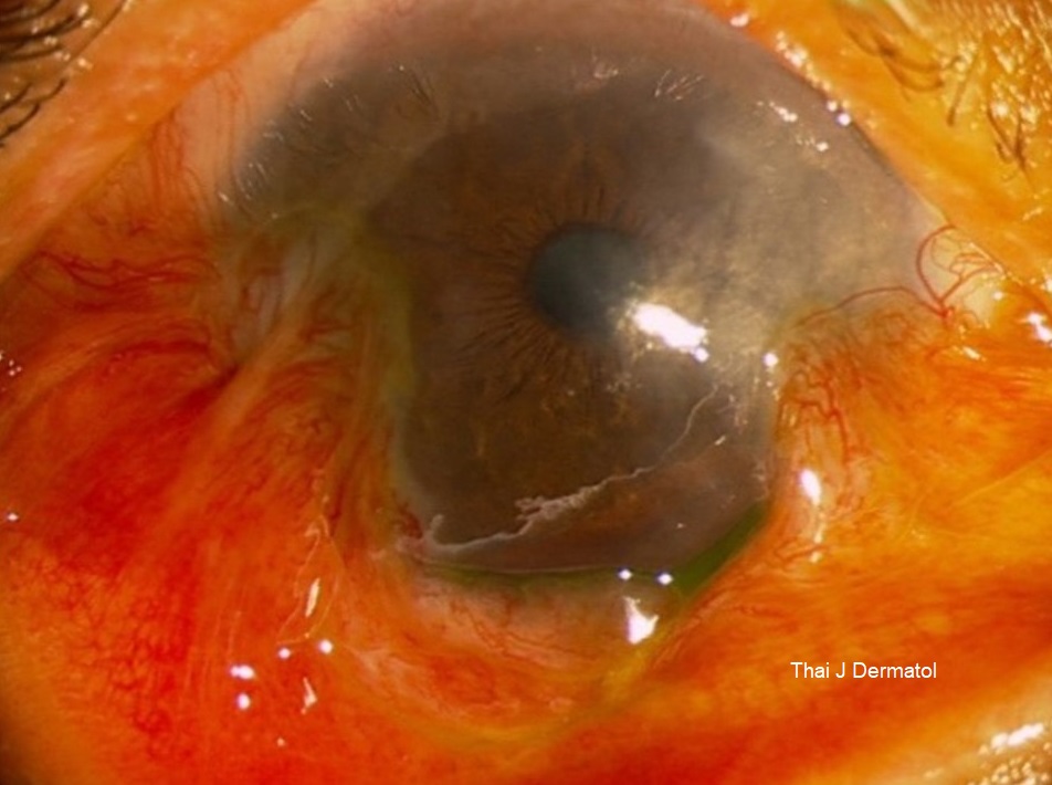
Ophthalmic involvement in autoimmune bullous diseases (AIBDs) can lead to permanent visual loss and blindness. Ocular manifestation usually parallel to active mucocutaneous disease. Apart from mucous membrane pemphigoid (MMP), pure ocular involvement in other AIBDs is exceedingly rare. We report three cases of AIBDs consisting of immunoglobulin (Ig) A/IgG pemphigus, linear IgA disease (LABD) and pemphigus vulgaris (PV) in which all patients presented with chronic cicatrizing conjunctivitis, mimicking MMP, without skin involvement. Our first patient’s conjunctival biopsy revealed separation of the stratified squamous epithelium with acantholysis. Direct immunofluorescence (DIF) demonstrated intercellular IgA deposition. Indirect immunofluorescence (IIF) was also positive for intercellular IgA. IgG enzyme-linked immunosorbent assay was positive for Desmoglein-1 (Dsg1) and negative for Anti-Dsg3. The diagnosis of IgA/IgG pemphigus was made. The second patient’s conjunctival biopsy showed fibrosis, chronic inflammation at subepithelial junction with separation. DIF revealed linear basement membrane IgA, leading to the diagnosis of LABD.
Conjunctival biopsy of the final case showed chronic conjunctivitis with focal suprabasal separation of the stratified squamous epithelium. Immunoperoxidase showed intercellular IgG and C3. IIF was positive for IgG. The final diagnosis was ocular PV. All patients received systemic corticosteroids together with azathioprine or dapsone with satisfactory results. To the best of our knowledge, this is the first reported case for ocular IgA/IgG pemphigus. The presence pure ocular involvement is extremely rare for LABD and PV. We emphasize that AIBDs exclusive to the eye is a diagnostic dilemma. Early recognition and treatment can halt the progression and prevent permanent visual loss.
References
2. Laforest C, Huilgol SC, Casson R, Selva D, Leibovitch I. Autoimmune bullous diseases: ocular manifestations and management. Drugs 2005; 65: 1767-79.
3. Talhari C, Althaus C, Megahed M. Ocular linear IgA disease resulting in blindness. Arch Dermatol 2006; 142: 786-7.
4. Letko E, Bhol K, Foster CS, Ahmed AR. Linear IgA bullous disease limited to the eye: a diagnostic dilemma: response to intravenous immunoglobulin therapy. Ophthalmology 2000; 107: 1524-8.
5. Daoud YJ, Cervantes R, Foster CS , Ahmed AR. Ocular pemphigus. J Am Acad Dermatol 2005; 53: 585-90.
6. Nishikawa T, Hashimoto T. Dermatoses with intraepidermal IgA deposits. Clin Dermatol 2000; 18: 315-8.
7. Bruckner AL, Fitzpatrick JE, Hashimoto T, Weston WL, Morelli JG. Atypical IgA/IgG pemphigus involving the skin, oral mucosa, and colon in a child: a novel variant of IgA pemphigus? Pediatr Dermatol 2005; 22: 321-7.
8. Teraki Y, Amagai N, Hashimoto T, Kusunoki T , Nishikawa T. Intercellular IgA dermatosis of childhood. Selective deposition of monomer IgA1 in the intercellular space of the epidermis. Arch Dermatol 1991; 127: 221-4.
9. Robinson ND, Hashimoto T, Amagai M , Chan LS. The new pemphigus variants. J Am Acad Dermatol 1999; 40: 649-71.
10. Heng A, Nwaneshiudu A, Hashimoto T, Amagai M , Stanley JR. Intraepidermal neutrophilic IgA/IgG antidesmocollin 1 pemphigus. Br J Dermatol 2006; 154: 1018-20.
11. Amagai M. Autoimmunity against desmosomal cadherins in pemphigus. J Dermatol Sci 1999; 20: 92-102.
12. Gooptu C, Mendelsohn S, Amagai M, Hashimoto T, Nishikawa T, Wojnarowska F. Unique immunobullous disease in a child with a predominantly IgA response to three desmosomal proteins. Br J Dermatol 1999; 141: 882-6.
13. Toosi S, Collins JW, Lohse CM, et al. Clinicopathologic features of IgG/IgA pemphigus in comparison with classic (IgG) and IgA pemphigus. Int J Dermatol 2016; 55: e184-90.
14. Peters MS, Rogers RS. Clinical correlations of linear IgA deposition at the cutaneous basement membrane zone. J Am Acad Dermatol 1989; 20: 761-70.
15. España A, Iranzo P, Herrero-González J, Mascaro JM Jr, Suárez R. Ocular involvement in pemphigus vulgaris - a retrospective study of a large Spanish cohort. J Dtsch Dermatol Ges 2017; 15: 396-403.
16. Hodak E, Kremer I, David M, et al. Conjunctival involvement in pemphigus vulgaris: a clinical, histopathological and immunofluorescence study. Br J Dermatol 1990; 123: 615-20.
Downloads
Published
How to Cite
Issue
Section
License
เนื้อหาและข้อมูลในบทความที่ลงตีพิมพ์ในวารสารโรคผิวหนัง ถือเป็นข้อคิดเห็นและความรับผิดชอบของผู้เขียนบทความโดยตรงซึ่งกองบรรณาธิการวารสาร ไม่จำเป็นต้องเห็นด้วย หรือร่วมรับผิดชอบใดๆ
บทความ ข้อมูล เนื้อหา รูปภาพ ฯลฯ ที่ได้รับการตีพิมพ์ในวารสารโรคผิวหนัง ถือเป็นลิขสิทธิ์ของวารสารฯ หากบุคคลหรือหน่วยงานใดต้องการนำทั้งหมดหรือส่วนหนึ่งส่วนใดไปเผยแพร่ต่อหรือเพื่อกระทำการใดๆ จะต้องได้รับอนุญาตเป็นลายลักอักษรจากบรรณาธิการวารสารโรคผิวหนังก่อนเท่านั้น

