
An atypical presentation of subcutaneous panniculitis-like T-Cell lymphoma
Keywords:
Facial swelling, Subcutaneous Panniculitis-like T-Cell Lymphoma (SPTL)Abstract
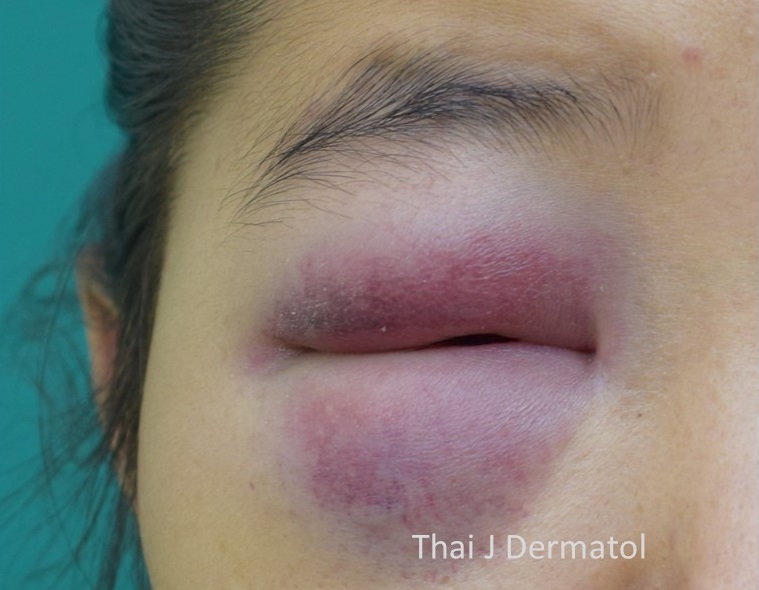
Subcutaneous panniculitis-like T-Cell Lymphoma (SPTL) is a rare cytotoxic T-cell lymphoma primarily localized to the subcutaneous tissue. Clinical presentations are usually consisted of painless indurated subcutaneous nodules or plaques with variable number and sizes. The most common sites of presentation are extremity and trunk. We reported a case of SPTL patient who presented with progressive painless facial and periorbital swelling with B symptoms.
References
Swerdlow SH, Campo E, Pileri SA, et al. The 2016 revision of the World Health Organization classification of lymphoid neoplasms. Blood 2016; 127: 2375-90.
López-Lerma I, Peñate Y, Gallardo F, et al. Subcutaneous panniculitis-like T-cell lymphoma: Clinical features, therapeutic approach, and outcome in a case series of 16 patients. J Am Acad Dermatol 2018; 79: 892-8.
Kong YY, Dai B, Kong JC, et al. Subcutaneous panniculitis-like T-cell lymphoma: a clinicopathologic, immunophenotypic, and molecular study of 22 Asian cases according to WHO-EORTC classification. Am J Surg Pathol 2008; 32: 1495-502.
Rolinski M, Brown VL, Ratnavel R. Subcutaneous panniculitis-like Tcell lymphoma. J Eur Acad Dermatol Venereol 2010; 24: 241-2.
Hoque SR, Child FJ, Whittaker SJ, et al. Subcutaneous panniculitis-like T-cell lymphoma: a clinicopathological, immunophenotypic and molecular analysis of six patients. Br J Dermatol 2003; 148: 516-25.
Go RS, Wester SM. Immunophenotypic and molecular features, clinical outcomes, treatments, and prognostic factors associated with subcutaneous panniculitis-like T-cell lymphoma: a systematic analysis of 156 patients reported in the literature. Cancer 2004; 101: 1404-13.
Guenova E, Schanz S, Hoetzenecker W, et al. Systemic corticosteroids for subcutaneous panniculitis-like T-cell lymphoma. Br J Dermatol 2014; 171: 891-4.
Sakurai E, Satoh T, Akiko YA, et al. Subcutaneous panniculitis-like T-cell lymphoma (SPTCL) with hemophagocytosis (HPS): successful treatment using high-dose chemotherapy (BFM-NHL & ALL-90) and autologous peripheral blood stem cell transplantation. J Clin Exp Hematop 2013; 53: 135-40.
Bhojaraja MV, Kistampally PK, Udupa KS, Thomas J. Subcutaneous Panniculitis-Like T-Cell Lymphoma: A Rare Tumour. J Clin Diagn Res 2016; 10: OD29-30.
Chinello M, Naviglio S, Remotti D, Locasciulli A, Ventura A. Subcutaneous panniculitis-like T-cell lymphoma presenting with diffuse cutaneous edema in a 2-year-old child. J Pediatr Hematol Oncol 2015; 37: 329-30.
Lee WJ, Moon IJ, Won CH, et al. Facial swelling: an atypical presentation of cutaneous lymphoma. Int J Dermatol 2016; 55: e440-6.
Hashimoto R, Uchiyama M, Maeno T. Case report of subcutaneous panniculitis-like T-cell lymphoma complicated by eyelid swelling. BMC Ophthalmol 2016; 16: 117.
Asati DP, Ingle V, Joshi D, Tiwari A. Subcutaneous panniculitis-like T-cell lymphoma with macrophage activation syndrome treated by cyclosporine and prednisolone. Indian Dermatol Online J 2016; 7: 529-32.
Kosari F, Akbarzadeh Hosseini S. Local facial edema: a novel presentation of subcutaneous panniculitis-like T-cell lymphoma in a 30-year-old Iranian woman. Acta Med Iran 2014;52: 950-3.
Velez NF, Ishizawar RC, Dellaripa PF, et al. Full facial edema: a novel presentation of subcutaneous panniculitis-like T-cell lymphoma. J Clin Oncol 2012; 30: e233-6.
Downloads
Published
How to Cite
Issue
Section
License
เนื้อหาและข้อมูลในบทความที่ลงตีพิมพ์ในวารสารโรคผิวหนัง ถือเป็นข้อคิดเห็นและความรับผิดชอบของผู้เขียนบทความโดยตรงซึ่งกองบรรณาธิการวารสาร ไม่จำเป็นต้องเห็นด้วย หรือร่วมรับผิดชอบใดๆ
บทความ ข้อมูล เนื้อหา รูปภาพ ฯลฯ ที่ได้รับการตีพิมพ์ในวารสารโรคผิวหนัง ถือเป็นลิขสิทธิ์ของวารสารฯ หากบุคคลหรือหน่วยงานใดต้องการนำทั้งหมดหรือส่วนหนึ่งส่วนใดไปเผยแพร่ต่อหรือเพื่อกระทำการใดๆ จะต้องได้รับอนุญาตเป็นลายลักอักษรจากบรรณาธิการวารสารโรคผิวหนังก่อนเท่านั้น

