
Solitary mycosis fungoides treated with photochemotherapy: A case report
Keywords:
unilesional, solitary, mycosis fungoidesAbstract
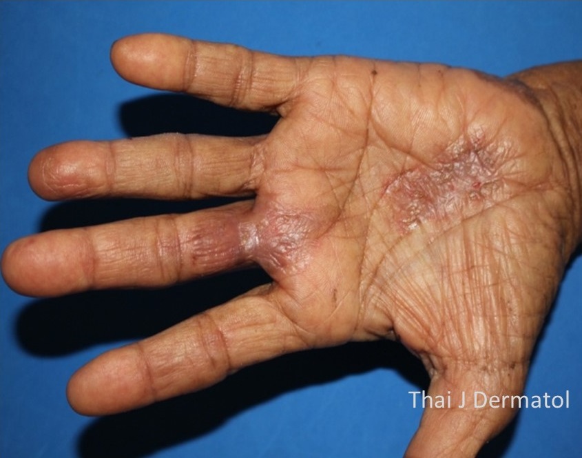
Mycosis fungoides (MF) is the most frequent diseases among cutaneous T-cell Lymphoma (CTCL). MF is categorized as being patch, plaque, or tumor stage, but patients may concurrently have more than one type of the lesion. Solitary or unilesional mycosis fungoides is a variant of MF characterized by a single lesion involving less than 5% of total body surface skin area. Clinical feature is single erythematous plaque occurs anywhere on the body such as scalp, face, trunk, upper and lower extremities. Histopathology is similar to MF which has atypical lymphoid infiltrate with epidermotropism, possible adnexal involvement. Many cases report presented relationship among MF, koebner phenomenon and cutaneous infection. However solitary MF usually has an indolent course and good prognosis. There are various therapeutic approaches to solitary MF such as radiotherapy, surgical excision, photodynamic therapy and topical corticosteroid. We report an 84-year-old woman presented with solitary scaly hyperkeratotic erythematous plaque with minimal erosions on left palm for 1 year. She had history of thorn injury at left palm before the lesion developed. The histopathological study, immunohistochemistry and T-cell receptor (TCR) gene rearrangement were compatible with MF. She was treated by topical psoralen plus UVA (PUVA) with resolved of lesion after 6 months of treatment without any side effect.
References
Ally MS, Robson A. A review of the solitary cutaneous T-cell lymphomas. J Cutan Pathol 2014; 41: 703-14.
Jackow CM, McHam JB, Friss A, Alvear J, Reveille JR, Duvic M. HLA-DR5 and DQB1*03 class II alleles are associated with cutaneous T-cell lymphoma. J Invest Dermatol 1996; 107: 373-6.
Mirvish JJ, Pomerantz RG, Falo LD JR et al. Role of infectious agents in cutaneous T-cell lymphoma: facts and controversies. Clin Dermatol 2013; 31: 423–31.
Abrams JT, Balin BJ, Vonderheid EC. Association between Sézary T cell-activating factor, Chlamydia pneumoniae, and cutaneous T cell lymphoma. Ann NY Acad Sci 2001; 941: 69–85.
Litvinov IV, Shtreis A, Kobayashi K, et al. Investigating potential exogenous tumor initiating and promoting factors for Cutaneous T-Cell Lymphomas (CTCL), a rare skin malignancy. Oncoimmunol 2016; 5: e1175799.
Slodownik D, Moshe S, Sprecher E, Goldberg I. Occupational mycosis fungoides – a case series. Int J Dermatol 2017; 56: 733–7.
Talpur R, Cox KM, Hu M, et al. Vitamin D deficiency in mycosis fungoides and Sezary syndrome patients is similar to other cancer patients. Clin Lymphoma Myeloma Leuk 2014; 14: 518–24.
Heald PW, Glusac EJ. Unilesional cutaneous T-cell lymphoma: clinical features, therapy, and follow-up of 10 patients with a treatment responsive mycosis fungoides variant. J Am Acad Dermatol 2000; 42: 283–5.
Ally MS, Pawade J, Tanaka M, et al. Solitary mycosis fungoides: a distinct clinicopathologic entity with a good prognosis: a series of 15 cases and literature review. J Am Acad Dermatol 2012; 67: 736-44.
Amitay-Laish I, Feinmesser M, Ben-Amitai D, et al. Unilesional folliculotropic mycosis fungoides: a unique variant of cutaneous lymphoma. J Eur Acad Dermatol Venereol 2016; 30: 25-9.
Downloads
Published
How to Cite
Issue
Section
License
เนื้อหาและข้อมูลในบทความที่ลงตีพิมพ์ในวารสารโรคผิวหนัง ถือเป็นข้อคิดเห็นและความรับผิดชอบของผู้เขียนบทความโดยตรงซึ่งกองบรรณาธิการวารสาร ไม่จำเป็นต้องเห็นด้วย หรือร่วมรับผิดชอบใดๆ
บทความ ข้อมูล เนื้อหา รูปภาพ ฯลฯ ที่ได้รับการตีพิมพ์ในวารสารโรคผิวหนัง ถือเป็นลิขสิทธิ์ของวารสารฯ หากบุคคลหรือหน่วยงานใดต้องการนำทั้งหมดหรือส่วนหนึ่งส่วนใดไปเผยแพร่ต่อหรือเพื่อกระทำการใดๆ จะต้องได้รับอนุญาตเป็นลายลักอักษรจากบรรณาธิการวารสารโรคผิวหนังก่อนเท่านั้น

