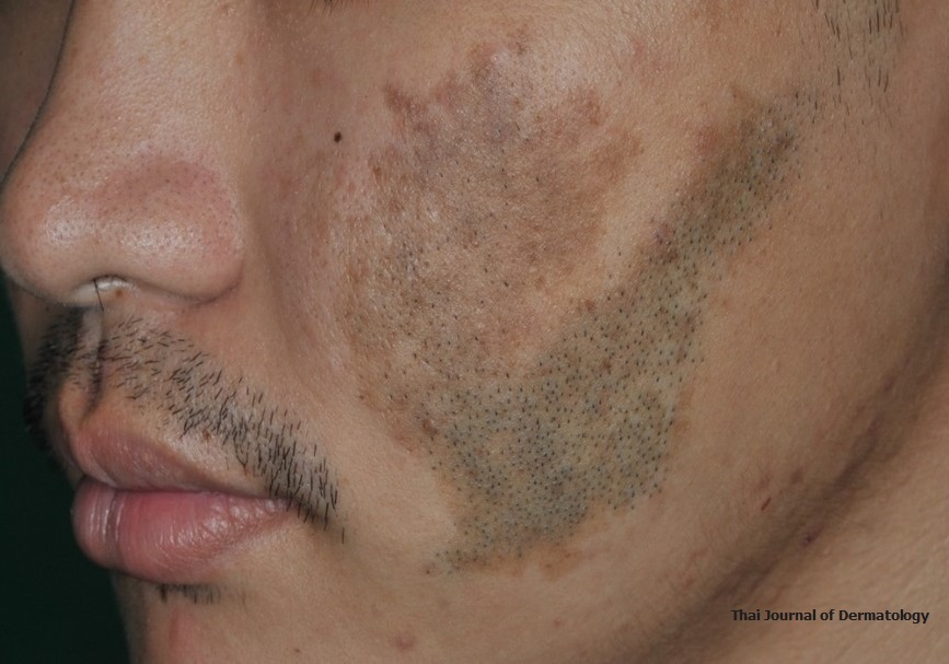
Giant congenital melanocytic nevus with Becker’s nevus-like presentation
Keywords:
congenital melanocytic nevus, giant congenital melanocytic nevusAbstract
KAMBHU NA AYUDHYA T, CHAWVAVANICH P. GIANT CONGENITAL MELANOCYTIC NEVUS WITH BECKER’S NEVUS-LIKE PRESENTATION. THAI J DERMATOL 2020;36:73-77.
INSTITUTE OF DERMATOLOGY, DEPARTMENT OF MEDICAL SERVICES, MINISTRY OF PUBLIC HEALTH, BANGKOK, THAILAND.
Congenital melanocytic nevus (CMN) is present at birth, clinically presents as a round brownish lesion with well-defined borders and hypertrichosis. The surface of the nevus may be slightly raised, papular, roughed, warty or cerebriform1,2. CMN is often composed of complex three histological patterns that include nevus cell type, neuroid type, and blue nevus cell type. In our case patient presented with hypertrichosis, flat hyperpigmented patches with an irregular border. From this clinical presentation, the differential diagnosis were Becker’s nevus, pigmented neurofibroma and congenital melanocytic nevus which can mimicking each other. Skin biopsy was showed tyrosinase-positive spindle cells. In this case, we conclude the diagnosis as congenital melanocytic nevus with neuroid type.
References
Kovalyshyn I, Braun R, Marghoob A. Congenital melanocytic naevi. Australas J Dermatol 2009;50:231-40.
Marghoob AA. Congenital melanocytic nevi. Evaluation and management. Dermatol Clin 2002;20:607-16.
Mizushima J, Nogita T, Higaki Y, Horikoshi T, Kawashima M. Dormant melanocytes in the dermis: do dermal melanocytes of acquired dermal melanocytosis exist from birth? Be J Dermatol 1998;139:349-50.
Rhodes AR. Congenital nevomelanocytic nevi: histologic patterns in the first year of life and evolution during childhood. Arch Dermatol 1986;122:1257-62.
Arneja JS, Gosain AK. Giant congenital melanocytic nevi. Plast Reconstr Surg 2009;124:1e-13e.
Kinsler VA, Birley J, Atherton DJ. Great Ormond Street Hospital for Children Registry for Congenital melanocytic naevi: prospective study 1988-2007. Part 2--Evaluation of treatments. Br J Dermatol 2009;160:387-92.
Slutsky JB, Barr JM, Femia A, Marghoob AA. Large congenital melanocytic nevi: associated risks and management considerations. Semin Cutan Med Surq 2010;29:79-84.
Wu D, Wang M, Wang X, et al. Lack of BRAF(V600E) mutations in giant congenital melanocytic nevi in a Chinese population. The Am J Dermatopathol 2011;33:341-4.
Castilla EE, da Graca Dutra M, Orioli-Parreiras IM. Epidemiology of congenital pigmented naevi: I. Br J Dermatol 1981;104:307-15.
Walton RG, Jacobs AH, Cox AJ. Pigmented lesions in newborn infants. Br J Dermatol 1976;95:389-96.
Viana AC, Gontijo B, Bittencourt FV. Giant congenital melanocytic nevus. An Bras Dermatol 2013;88:863-78.
Balin SJ, Barnhill RL. Benign Melanocytic Neoplasms. In: Bolognia J, editor. Dermatology 4th edition: Elsevier 2018.p.1954-84.
Reed WB, Becker SW Sr, Becker SW Jr, Nickel WR. Giant pigmented nevi, melanoma, and leptomeningeal melanocytosis: a clinical and histopathological study. Arch Dermatol 1965;91:100-19.
From L. Congenital Nevi-Let's Be Practical. Pediatr Dermatol 1992;9:345-6.
Mark GJ, Mihm MC, Liteplo MG, Reed RJ, Clark WH. Congenital melanocytic nevi of the small and garment type. Clinical, histologic, and ultrastructural studies. Hum Pathol 1973;4:395-418.
Fetsch JF, Michal M, Miettinen M. Pigmented (melanotic) neurofibroma: a clinicopathologic and immunohistochemical analysis of 19 lesions from 17 patients. Am J Surq Pathol 2000;24:331-43.

Downloads
Published
How to Cite
Issue
Section
License
เนื้อหาและข้อมูลในบทความที่ลงตีพิมพ์ในวารสารโรคผิวหนัง ถือเป็นข้อคิดเห็นและความรับผิดชอบของผู้เขียนบทความโดยตรงซึ่งกองบรรณาธิการวารสาร ไม่จำเป็นต้องเห็นด้วย หรือร่วมรับผิดชอบใดๆ
บทความ ข้อมูล เนื้อหา รูปภาพ ฯลฯ ที่ได้รับการตีพิมพ์ในวารสารโรคผิวหนัง ถือเป็นลิขสิทธิ์ของวารสารฯ หากบุคคลหรือหน่วยงานใดต้องการนำทั้งหมดหรือส่วนหนึ่งส่วนใดไปเผยแพร่ต่อหรือเพื่อกระทำการใดๆ จะต้องได้รับอนุญาตเป็นลายลักอักษรจากบรรณาธิการวารสารโรคผิวหนังก่อนเท่านั้น


