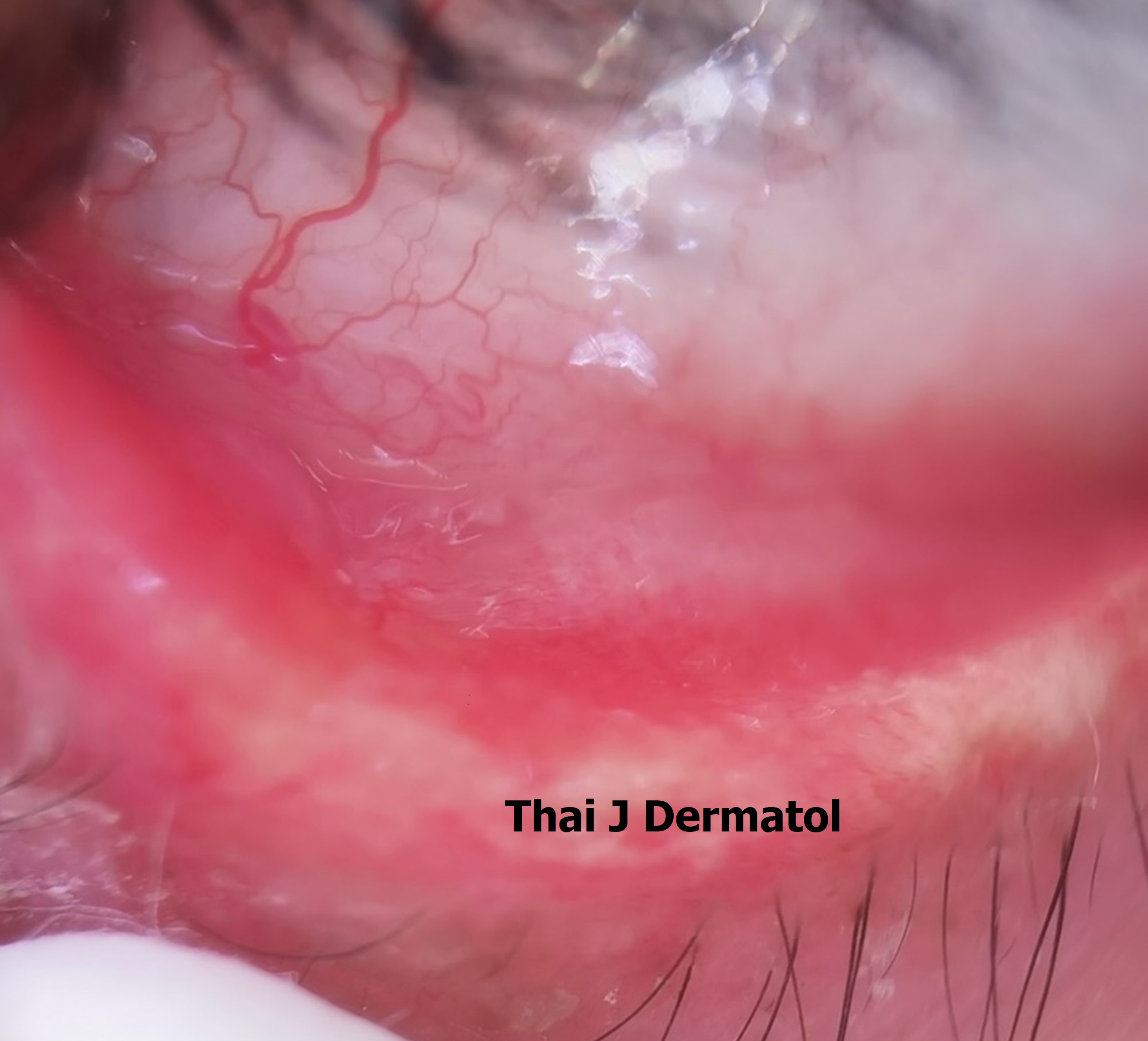
Eyelid Discoid Lupus Erythematosus Presenting with Chronic Blepharoconjunctivitis, Mimicking Mucous Membrane Pemphigoid
Keywords:
Blepharoconjunctivitis, dermoscopy, discoid lupus erythematousAbstract
Discoid lupus erythematosus (DLE) is the most common form of chronic cutaneous lupus erythematosus. DLE can occur on sun-exposed areas and mucosal surfaces. However, DLE on the ocular mucosa, presenting as chronic blepharoconjunctivitis, is rare and may lead to misdiagnosis with other skin conditions, especially autoimmune diseases like mucous membrane pemphigoid. Moreover, due to the rarity of DLE on the eye, the delay in diagnosis may result in complications and morbidity on the effected eyes. Dermoscopy is a non-invasive tool, commonly used to aid the diagnosis of many skin conditions. Dermoscopic findings in DLE are well-established. However, in this case we used dermoscopy to examine DLE lesions on the ocular and eyelid skin surface which is rarely reported in the literature. Our dermoscopic evaluation showed telangiectasia, non-atrophic round erythema, brownish pigmentation with adherent scales on skin surface and non-atrophic round erythema with adherent scales on mucosal surface. Antimalarial drugs are the first-line systemic therapy and also most commonly used in the literatures for eyelids DLE. In this case, we also reported that dapsone in addition to hydroxychloroquine were effective treatments for eyelid DLE. Moreover, multidisciplinary care with ophthalmologist is essential to maximize treatment outcome and prevent further complications.
References
Okon LG, Werth VP. Cutaneous lupus erythematosus: diagnosis and treatment. Best Pract Res Clin Rheumatol 2013;27:391-404.
Laforest C, Huilgol SC, Casson R, Selva D, Leibovitch I. Autoimmune bullous diseases: ocular manifestations and management. Drugs 2005;65:1767-79.
Amescua G, Akpek EK, Farid M, et al. Blepharitis Preferred Practice Pattern®. Ophthalmology 2019;126:P56-93.
Chomiciene A, Stankeviciute R, Malinauskiene L, et al. Rare cause of periorbital and eyelids lesions: Discoid lupus erythematosus misdiagnosed as allergy. Ann Allergy Asthma Immunol 2017;119:568-9.
Ghauri AJ, Valenzuela AA, O'Donnell B, Selva D, Madge SN. Periorbital Discoid Lupus Erythematosus. Ophthalmology 2012;119:2193-4.e11.
Kopsachilis N, Tsaousis KT, Tourtas T, Tsinopoulos IT. Severe chronic blepharitis and scarring ectropion associated with discoid lupus erythematosus. Clin Exp Optom 2013;96:124-5.
Arrico L, Abbouda A, Abicca I, Malagola R. Ocular Complications in Cutaneous Lupus Erythematosus: A Systematic Review with a Meta-Analysis of Reported Cases. J Ophthalmol 2015;2015:254260.
Wu MY, Wang CH, Ng CY, et al. Periorbital erythema and swelling as a presenting sign of lupus erythematosus in tertiary referral centers and literature review. Lupus 2018;27:1828-37.
Lallas A, Apalla Z, Lefaki I, et al. Dermoscopy of discoid lupus erythematosus. Br J Dermatol 2013;168:284-8.
Salah E. Clinical and dermoscopic spectrum of discoid lupus erythematosus: novel observations from lips and oral mucosa. Int J Dermatol 2018;57:830-6.
Costamilan LZ, Cerci FB, Werner B. Erythematous Plaque on the Inferior Eyelid. JAMA Dermatol 2018;154:957-8.

Downloads
Published
How to Cite
Issue
Section
License
เนื้อหาและข้อมูลในบทความที่ลงตีพิมพ์ในวารสารโรคผิวหนัง ถือเป็นข้อคิดเห็นและความรับผิดชอบของผู้เขียนบทความโดยตรงซึ่งกองบรรณาธิการวารสาร ไม่จำเป็นต้องเห็นด้วย หรือร่วมรับผิดชอบใดๆ
บทความ ข้อมูล เนื้อหา รูปภาพ ฯลฯ ที่ได้รับการตีพิมพ์ในวารสารโรคผิวหนัง ถือเป็นลิขสิทธิ์ของวารสารฯ หากบุคคลหรือหน่วยงานใดต้องการนำทั้งหมดหรือส่วนหนึ่งส่วนใดไปเผยแพร่ต่อหรือเพื่อกระทำการใดๆ จะต้องได้รับอนุญาตเป็นลายลักอักษรจากบรรณาธิการวารสารโรคผิวหนังก่อนเท่านั้น


