
Hypopigmented Lesions: An Unusual Initial Presentation of Infantile Langerhans Cell Histiocytosis
Keywords:
Langerhans cell histiocytosis, LCH, hypopigmentationAbstract
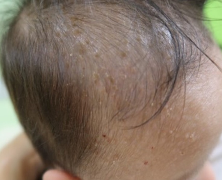
Langerhans cell histiocytosis is a rare neoplasm. Patients are typically presented with small, translucent, yellowish crusted papules on the trunk, intertriginous areas, and scalp. However, previous literature has reported hypopigmented lesions as an unusual cutaneous presentation, emphasizing the need for clinician awareness to aid a prompt diagnosis. Herein, we present a case of langerhans cell histiocytosis, whose cutaneous presentations were multiple hypopigmented lesions and yellowish crusts–an uncommon manifestation of langerhans cell histiocytosis.
References
Allen CE, Merad M, McClain KL. Langerhans-Cell Histiocytosis. N Engl J Med 2018;379:856-68.
Ribeiro KB, Degar B, Antoneli CB, Rollins B, Rodriguez-Galindo C. Ethnicity, race, and socioeconomic status influence incidence of Langerhans cell histiocytosis. Pediatr Blood Cancer 2015;62:982-7.
Poompuen S, Chaiyarit J, Techasatian L. Diverse cutaneous manifestation of Langerhans cell histiocytosis: a 10-year retrospective cohort study. Eur J Pediatr 2019;178:771-6.
Stein SL, Paller AS, Haut PR, Mancini AJ. Langerhans cell histiocytosis presenting in the neonatal period: a retrospective case series. Arch Pediatr Adolesc Med 2001;155:778-83.
Haupt R, Minkov M, Astigarraga I, et al. Langerhans cell histiocytosis (LCH): guidelines for diagnosis, clinical work-up, and treatment for patients till the age of 18 years. Pediatr Blood Cancer 2013;60:175-84.
Mehta B, Amladi S. Langerhans cell histiocytosis presenting as hypopigmented papules. Pediatr Dermatol 2010;27:215-7.
Krooks J, Minkov M, Weatherall AG. Langerhans cell histiocytosis in children: History, classification, pathobiology, clinical manifestations, and prognosis. J Am Acad Dermatol 2018;78:1035-44.
Feroze K, Unni M, Jayasree MG, Seethalekshmy NV. Langerhans cell histiocytosis presenting with hypopigmented macules. Indian J Dermatol Venereol Leprol 2008;74:670-2.
Kaddu S, Mulyowa G, Kovarik C. Hypopigmented scaly, scalp and facial lesions and disfiguring exopthalmus. Langerhans cell histiocytosis (LCH). Clin Exp Dermatol 2010;35:e52-3.
Mori S, Adar T, Kazlouskaya V, Alexander JB, Heilman E, Glick SA. Cutaneous Langerhans cell histiocytosis presenting with hypopigmented lesions: Report of two cases and review of literature. Pediatr Dermatol 2018;35:502-6.
Lozano Masdemont B, Gómez-Recuero Muñoz L, Villanueva Álvarez-Santullano A, Parra Blanco V, Campos Domínguez M. Langerhans cell histiocytosis mimicking lichen nitidus with bone involvement. Australas J Dermatol 2017;58:231-3.
Longaker MA, Frieden IJ, LeBoit PE, Sherertz EF. Congenital "self-healing" Langerhans cell histiocytosis: the need for long-term follow-up. J Am Acad Dermatol 1994;31:910-6.
Parimi LR, You J, Hong L, Zhang F. Congenital self-healing reticulohistiocytosis with spontaneous regression. An Bras Dermatol 2017;92:553-5.
Uaratanawong R, Kootiratrakarn T, Sudtikoonaseth P, Issara A, Kattipathanapong P. Congenital self-healing reticulohistiocytosis presented with multiple hypopigmented flat-topped papules: a case report and review of literatures. J Med Assoc Thai 2014;97:993-7.
Battistella M, Fraitag S, Teillac DH, Brousse N, de Prost Y, Bodemer C. Neonatal and early infantile cutaneous langerhans cell histiocytosis: comparison of self-regressive and non-self-regressive forms. Arch Dermatol 2010;146:149-56.
Downloads
Published
How to Cite
Issue
Section
License
Copyright (c) 2022 Thai Journal of Dermatology

This work is licensed under a Creative Commons Attribution-NonCommercial-NoDerivatives 4.0 International License.
เนื้อหาและข้อมูลในบทความที่ลงตีพิมพ์ในวารสารโรคผิวหนัง ถือเป็นข้อคิดเห็นและความรับผิดชอบของผู้เขียนบทความโดยตรงซึ่งกองบรรณาธิการวารสาร ไม่จำเป็นต้องเห็นด้วย หรือร่วมรับผิดชอบใดๆ
บทความ ข้อมูล เนื้อหา รูปภาพ ฯลฯ ที่ได้รับการตีพิมพ์ในวารสารโรคผิวหนัง ถือเป็นลิขสิทธิ์ของวารสารฯ หากบุคคลหรือหน่วยงานใดต้องการนำทั้งหมดหรือส่วนหนึ่งส่วนใดไปเผยแพร่ต่อหรือเพื่อกระทำการใดๆ จะต้องได้รับอนุญาตเป็นลายลักอักษรจากบรรณาธิการวารสารโรคผิวหนังก่อนเท่านั้น

