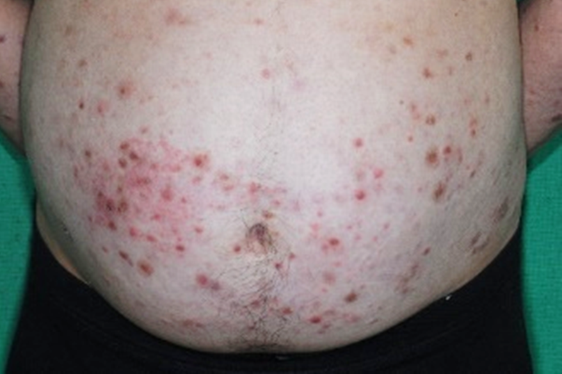
A Case Report of Unrecognized Lepromatous Leprosy Occurring in Conjunction with Pemphigus Vulgaris
คำสำคัญ:
Lepromatous leprosy, Pemphigus vulgaris, Oral immunosuppressive agentsบทคัดย่อ
Leprosy is a chronic, infectious disease of a significant public health concern, primarily affecting the skin and peripheral nerves. The causative agent of this disease is Mycobacterium leprae, and despite its recognition as an ancient affliction, it continues to be occasionally encountered in the southern regions of Thailand. The manifestation and progression of leprosy are known to be influenced by the host's genetic background and immune response. We present a case of a 45-year-old Thai male who developed new infiltrative papules and nodules while undergoing oral immunosuppressive therapy for the treatment of his underlying pemphigus vulgaris condition. Upon histopathological examination, the presence of histiocytes containing abundant acid-fast bacilli was observed. The patient was successfully treated through a 2-year course of multidrug therapy. This case highlights the challenges in diagnosing leprosy in immunocompromised individuals and underscores the importance of vigilant recognition of the disease by healthcare practitioners. It is imperative that medical professionals remain aware of the potential for leprosy to present in unexpected ways and take necessary steps to diagnose and treat the disease effectively.
เอกสารอ้างอิง
Maymone MB, Laughter M, Venkatesh S, et al. Leprosy: Clinical aspects and diagnostic techniques J Am Acad Dermatol 2020;83:1-14.
Nath I, Saini C, Valluri VL. Immunology of leprosy and diagnostic challenges. Clin in Dermatol 2015;33:90-8.
Reibel F, Cambau E, Aubry A. Update on the epidemiology, diagnosis, and treatment of leprosy. Med Mal Infect 2015;45:383-93.
Sadhu S, Mitra DK. Emerging Concepts of Adaptive Immunity in Leprosy. Front Immunol 2018;9:604.
da Silva MB, Portela JM, Li W, et al. Evidence of zoonotic leprosy in Pará, Brazilian Amazon, and risks associated with human contact or consumption of armadillos. PLoS Negl Trop Dis 2018;12:e0006532.
Moet FJ, Pahan D, Schuring RP, Oskam L, Richardus JH. Physical distance, genetic relationship, age, and leprosy classification are independent risk factors for leprosy in contacts of patients with leprosy. J Infect Dis 20061;193:346-53.
Trindade MAB, Palermo MdL, Pagliari C, et al. Leprosy in transplant recipients: report of a case after liver transplantation and review of the literature. Transpl Infect Dis 2011;13:63-9.
Aarestrup FM, Sampaio EP, de Moraes MO, Albuquerque EC, Castro AP, Sarno EN. Experimental Mycobacterium leprae infection in BALB/c mice: effect of BCG administration on TNF-alpha production and granuloma development. Int J Lepr Other Mycobact Dis 2000;68:156-66.
Barroso DH, Brandão JG, Andrade ESN, et al. Leprosy detection rate in patients under immunosuppression for the treatment of dermatological, rheumatological, and gastroenterological diseases: a systematic review of the literature and meta-analysis. BMC Infect Dis 2021;21:1-9.
Leshem YA, Gdalevich M, Ziv M, David M, Hodak E, Mimouni D. Opportunistic infections in patients with pemphigus. J Am Acad Dermatol 2014;71:284-92.

ดาวน์โหลด
เผยแพร่แล้ว
เวอร์ชัน
- 2023-09-11 (2)
- 2023-09-04 (1)
รูปแบบการอ้างอิง
ฉบับ
ประเภทบทความ
สัญญาอนุญาต
ลิขสิทธิ์ (c) 2023 วารสารโรคผิวหนัง

อนุญาตภายใต้เงื่อนไข Creative Commons Attribution-NonCommercial-NoDerivatives 4.0 International License.
เนื้อหาและข้อมูลในบทความที่ลงตีพิมพ์ในวารสารโรคผิวหนัง ถือเป็นข้อคิดเห็นและความรับผิดชอบของผู้เขียนบทความโดยตรงซึ่งกองบรรณาธิการวารสาร ไม่จำเป็นต้องเห็นด้วย หรือร่วมรับผิดชอบใดๆ
บทความ ข้อมูล เนื้อหา รูปภาพ ฯลฯ ที่ได้รับการตีพิมพ์ในวารสารโรคผิวหนัง ถือเป็นลิขสิทธิ์ของวารสารฯ หากบุคคลหรือหน่วยงานใดต้องการนำทั้งหมดหรือส่วนหนึ่งส่วนใดไปเผยแพร่ต่อหรือเพื่อกระทำการใดๆ จะต้องได้รับอนุญาตเป็นลายลักอักษรจากบรรณาธิการวารสารโรคผิวหนังก่อนเท่านั้น

