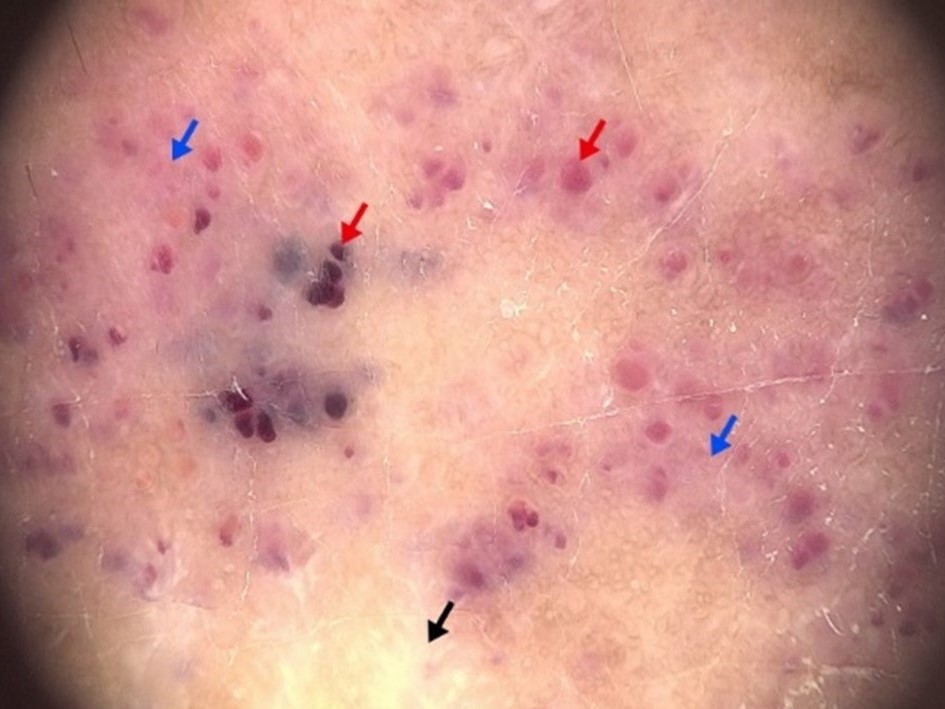
Non-targetoid Hobnail Hemangioma: Two Case Reports and Literature Review
Keywords:
Hobnail hemangioma, Targetoid hemosiderotic hemangioma, Superficial hemosiderotic lymphovascular malformation, DermoscopyAbstract
Hobnail hemangioma (HH) or targetoid hemosiderotic hemangioma is a benign vascular tumor with classic presentation of pale and ecchymotic halo. However, there are diversity in clinical and dermoscopic manifestations of HH, which can make diagnosis challenging in cases where the characteristic targetoid appearance is absent. This report discussed two cases of non-targetoid HH. A 15-year-old Thai female presented with erythematous plaque on her left knee, where dermoscopic findings include red and dark lacunae with white structures and a reddish homogeneous area, which are indicative of HH. The second case involves a 67-year-old Caucasian male presented with an ulcerated crusted nodule on his upper back. In this case, histopathological examination was necessary to rule out skin cancer and confirm the diagnosis of HH. This study provides an overview of HH and the challenges posed by non-targetoid presentations, emphasizing the importance of clinical and dermoscopic evaluation, as well as histopathological examination in uncertain cases.
References
Santa Cruz DJ, Aronberg J. Targetoid hemosiderotic hemangioma. J Am Acad Dermatol 1988;19:550-8.
Joyce JC, Keith PJ, Szabo S, Holland KE. Superficial hemosiderotic lymphovascular malformation (hobnail hemangioma): a report of six cases. Pediatr Dermatol 2014;31:281-5.
Porriño-Bustamante ML, Aneiros-Fernández J, Retámero JA, Fernández-Pugnaire MA. Hobnail Hemangioma With an Unusual Clinical Presentation. J Cutan Med Surg 2017;21:164-6.
Trindade F, Kutzner H, Tellechea Ó, Requena L, Colmenero I. Hobnail hemangioma reclassified as superficial lymphatic malformation: a study of 52 cases. J Am Acad Dermatol 2012;66:112-5.
Yoon SY, Kwon HH, Jeon HC, Lee JH, Cho S. Congenital and multiple hobnail hemangiomas. Ann Dermatol 2011;23:539-43.
Zaballos P, Llambrich A, Del Pozo LJ, et al. Dermoscopy of Targetoid Hemosiderotic Hemangioma: A Morphological Study of 35 Cases. Dermatology 2015;231:339-44.
Behera B, Chandrashekar L, Thappa DM, Gochhait D, Ayyanar P. Atypical clinical and dermoscopic features of hobnail hemangioma. Indian J Dermatol Venereol Leprol 2021;87:93-7.
Papageorgiou V, Apalla Z, Sotiriou E, et al. The limitations of dermoscopy: false-positive and false-negative tumours. J Eur Acad Dermatol Venereol 2018;32:879-88.
Enei ML, Paschoal FM, Valdes R. Arborizing vessels in a targetoid hemosiderotic hemangioma: mistaken dermoscopic diagnosis of basal cell carcinoma. Dermatol Pract Concept 2017;7:43-7.
Piccolo V, Russo T, Mascolo M, Staibano S, Baroni A. Dermoscopic misdiagnosis of melanoma in a patient with targetoid hemosiderotic hemangioma. J Am Acad Dermatol 2014;71:e179-81

Downloads
Published
How to Cite
Issue
Section
License
Copyright (c) 2024 Thai Journal of Dermatology

This work is licensed under a Creative Commons Attribution-NonCommercial-NoDerivatives 4.0 International License.
เนื้อหาและข้อมูลในบทความที่ลงตีพิมพ์ในวารสารโรคผิวหนัง ถือเป็นข้อคิดเห็นและความรับผิดชอบของผู้เขียนบทความโดยตรงซึ่งกองบรรณาธิการวารสาร ไม่จำเป็นต้องเห็นด้วย หรือร่วมรับผิดชอบใดๆ
บทความ ข้อมูล เนื้อหา รูปภาพ ฯลฯ ที่ได้รับการตีพิมพ์ในวารสารโรคผิวหนัง ถือเป็นลิขสิทธิ์ของวารสารฯ หากบุคคลหรือหน่วยงานใดต้องการนำทั้งหมดหรือส่วนหนึ่งส่วนใดไปเผยแพร่ต่อหรือเพื่อกระทำการใดๆ จะต้องได้รับอนุญาตเป็นลายลักอักษรจากบรรณาธิการวารสารโรคผิวหนังก่อนเท่านั้น


