
Extradigital Glomus Tumor: A Case Report of Pain on Back Over 10 Years
Keywords:
Glomus tumor, Extradigital glomus tumor, Painful cutaneous tumorAbstract
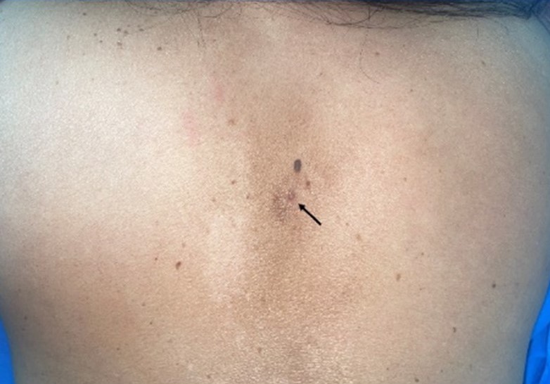
Glomus tumors are benign neoplasms of the glomus body, commonly found in the subungual area of digits. Occasionally, these tumors occur outside the digital region, making diagnosis more challenging. We report a case of 62-year-old woman who experienced pain on her back for 10 years. A careful examination revealed a small erythematous papule on the interscapular area. Dermoscopic findings showed homogenous, unstructured purplish area. A punch excision was performed, and histological and immunohistochemistry analysis of the tissue was consistent with a glomus tumor. The patient reported no pain following the procedure. This case report highlights the clinical, dermoscopic, and histological features of extradigital glomus tumors. It also emphasizes the importance of including these tumors in the differential diagnosis of painful nodules and demonstrates the effectiveness of complete surgical excision in achieving excellent outcomes.
References
Cohen PR. Glomus Extradigital Tumor: A Case Report of an Extradigital Glomus Tumor on the Wrist and Comprehensive Review of Glomus Tumors. Cureus 2023;15:e38737.
Chou T, Pan SC, Shieh SJ, Lee JW, Chiu HY, Ho CL. Glomus Tumor: Twenty-Year Experience and Literature Review. Ann Plast Surg 2016;76:S35-40.
Earley S. Vanilloid and melastatin transient receptor potential channels in vascular smooth muscle. Microcirculation 2010;17:237-49.
Abou Jaoude J, Farah AR, Sargi Z, Khairallah S, Fakih C. Glomus tumors: report on eleven cases and a review of the literature. Chirurgie de la Main 2000;19:243-52.
Álvarez-Salafranca M, Bañuls J, Thomas L, et al. Dermoscopy of glomus tumour: a cross-sectional study of 86 cases. J Eur Acad Dermatol Venereol. 2022;36:2016-24.
del Carpio GS, Burgos EMP, Kreilinger JJP, Taboada DB, Pensado MP, Viñé MT. Case series of extradigital glomus tumors: imaging findings, differential diagnosis and radiologic–pathologic correlation. Egyptian Journal of Radiology and Nuclear Medicine 2024;55:10.
Al-Qattan MM, Al-Namla A, Al-Thunayan A, Al-Subhi F, El-Shayeb AF. Magnetic resonance imaging in the diagnosis of glomus tumours of the hand. J Hand Surg Br 2005;30:535-40.
Miyamoto H, Wada H. Localized multiple glomangiomas on the foot. J Dermatol 2009;36:604-7.
Wu RC, Gao YH, Sun WW, Zhang XY, Zhang SP. Glomangiomatosis - immunohistochemical study: A case report. World J Clin Cases 2022;10:5406-13.
Folpe AL, Fanburg-Smith JC, Miettinen M, Weiss SW. Atypical and malignant glomus tumors: analysis of 52 cases, with a proposal for the reclassification of glomus tumors. Am J Surg Pathol 2001;25:1-12.
Downloads
Published
How to Cite
Issue
Section
License
Copyright (c) 2025 Thai Journal of Dermatology

This work is licensed under a Creative Commons Attribution-NonCommercial-NoDerivatives 4.0 International License.
เนื้อหาและข้อมูลในบทความที่ลงตีพิมพ์ในวารสารโรคผิวหนัง ถือเป็นข้อคิดเห็นและความรับผิดชอบของผู้เขียนบทความโดยตรงซึ่งกองบรรณาธิการวารสาร ไม่จำเป็นต้องเห็นด้วย หรือร่วมรับผิดชอบใดๆ
บทความ ข้อมูล เนื้อหา รูปภาพ ฯลฯ ที่ได้รับการตีพิมพ์ในวารสารโรคผิวหนัง ถือเป็นลิขสิทธิ์ของวารสารฯ หากบุคคลหรือหน่วยงานใดต้องการนำทั้งหมดหรือส่วนหนึ่งส่วนใดไปเผยแพร่ต่อหรือเพื่อกระทำการใดๆ จะต้องได้รับอนุญาตเป็นลายลักอักษรจากบรรณาธิการวารสารโรคผิวหนังก่อนเท่านั้น

