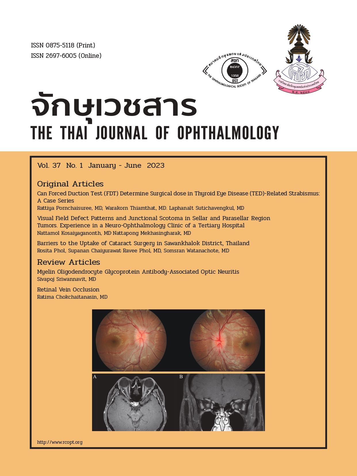
Retinal Vein Occlusion
Abstract
การอุดตันของหลอดเลือดดำจอตา (Retinal vein occlusion; RVO) เป็นหนึ่งในสาเหตุสำคัญของกลุ่มโรคหลอดเลือดจอตา ที่ทำให้เกิดการสูญเสียการมองเห็นเฉียบพลันในผู้สูงอายุ โดยพบได้เป็นอันดับสองรองลงมาจากภาวะเบาหวานขึ้นจอตา1 เเบ่งตามกายวิภาคออกเป็น 3 กลุ่มหลักๆ ได้แก่ หลอดเลือดดำที่จอตาอุดตัน (Central retinal vein occlusion; CRVO), แขนงหลอดเลือดดำที่จอตาอุดตัน (Branch retinal vein occlusion; BRVO) และหลอดเลือดดำที่จอตาอุดตันครึ่งซีก (Hemi retinal vein occlusion; HRVO) โดยในบทความนี้จะกล่าวถึง CRVO และ BRVO เป็นหลัก หลอดเลือดดำที่จอตาอุดตัน (CRVO) คือ ภาวะที่มีการอุดตันของหลอดเลือดดำหลักบริเวณด้านหลังแผ่น Lamina cribosa ของเส้นประสาทตา (optic nerve) มีอุบัติการณ์ของโรคโดยเฉลี่ย 0.5-0.8% แขนงหลอดเลือดดำที่จอตาอุดตัน (BRVO) พบได้บ่อยกว่า มีอุบัติการณ์ 0.44-1.6% แบ่งออกเป็น 2 กลุ่ม ได้แก่ major BRVO คือ มีการอุดตันของหลอดเลือดดำของจอตา 1 จตุภาค (quadrant) และ macular BRVO มีการอุดตันของหลอดเลือดดำจอตาภายในจุดภาพชัด (macula) โดยใน major BRVO พบได้บ่อยบริเวณ superotemporal area 58.1-66% และใน macular BRVO พบบริเวณ superior area 81%
References
Cugati S, Wang JJ, Rochtchina E, Mitchell P. Tenyear incidence of retinal vein occlusion in an older population: the Blue Mountains Eye Study: The blue mountains eye study. Arch Ophthalmol [Internet]. 2006;124(5):726–32. Available from: http://dx.doi.org/10.1001/archopht.124.5.726
Risk factors for central retinal vein occlusion. The Eye Disease Case-Control Study Group. Arch Ophthalmol. 1996;114(5):545-54.
Stephen JK, Amani F, Jaclyn LK, Shriji LK, Franco P, Lucia MR. Basic and Clinical Science Course:Retina and Vitreous. 2022.
Andrew PS, David PW, Srinivas RH, Peter RS, Ryan’s W. Ryan’s Retina. Retina.
Central Vein Occlusion Study (CVOS) [Internet]. Clinicaltrials.gov. [cited 2022 Oct 19]. Available from: https://clinicaltrials.gov/ct2/show/NCT00000131
Retinal vein occlusion during the early acute phase. Arbeitsphysiologie [Internet]. 1990;228(3):201–17. Available from: http://dx.doi.org/10.1007/
bf00920022
Prasad PS, Tsui I. Wide-field retinal imaging of branch retinal vein occlusions. Atlas of Wide-Field Retinal Angiography and Imaging. 2016: 69–81. Available at: https://doi.org/10.1007/978-3-319-17864-6_6.
An W, Zhao Q, Yu R, Han J. The role of optical coherence tomography angiography in distinguishing ischemic versus non-ischemic central retinal vein occlusion. BMC Ophthalmology. 2022;22(1):413. Available at: https://doi.org/10.1186/s12886-022-02637-y.
Central retinal vein occlusion [Internet]. Eyerounds. org. [cited 2022 Oct 19]. Available from: https://eyerounds.org/article/CRVO/
Morris R. Retinal vein occlusion. Kerala j ophthalmol [Internet]. 2016 [cited 2022 Oct 19];28(1):4. Available from: https://www.kjophthal.com/article.asp?issn=0976-6677;year=2016;volume=28;issue=1;spage=4;epage=13;aulast=Morris
Li C, Wang R, Liu G, Ge Z, Jin D, Ma Y, et al. Efficacy of panretinal laser in ischemic central retinal vein occlusion: A systematic review. Exp Ther Med [Internet]. 2019;17(1):901–10. Available from: http://dx.doi.org/10.3892/etm.2018.7034
Evaluation of grid pattern photocoagulation for macular edema in central vein occlusion. The Central Vein Occlusion Study Group M report.
Ophthalmology [Internet]. 1995;102(10):1425–33. Available from: http://dx.doi.org/10.1016/s0161-6420(95)30849-4
occlusion. Available at: https://www.cochrane.org/CD009510/EYES_anti-vascular-endothelial-growthfactor-macular-oedema-secondary-branch-retinal-vein-occlusion (Accessed: March 17, 2023).
Vander JF. Central and Hemicentral retinal vein occlusion: Role of anti–platelet aggregation agents and anticoagulants. Yearbook of Ophthalmology, 2012:130–1. Available at: https://doi.org/10.1016/j. yoph.2011.12.006.
Thomas GN. et al. Central retinal vein occlusion: The effect of antiplatelet and anticoagulant agents. Journal of VitreoRetinal Diseases. 2021;6(2):97-103. Available at: https://doi.org/10.1177/24741264211028508.
Downloads
Published
Issue
Section
License
Copyright (c) 2023 THE THAI JOURNAL OF OPHTHALMOLOGY

This work is licensed under a Creative Commons Attribution-NonCommercial-NoDerivatives 4.0 International License.
The Thai Journal of Ophthalmology (TJO) is a peer-reviewed, scientific journal published biannually for the Royal College of Ophthalmologists of Thailand. The objectives of the journal is to provide up to date scientific knowledge in the field of ophthalmology, provide ophthalmologists with continuing education, promote cooperation, and sharing of opinion among readers.
The copyright of the published article belongs to the Thai Journal of Ophthalmology. However the content, ideas and the opinions in the article are from the author(s). The editorial board does not have to agree with the authors’ ideas and opinions.
The authors or readers may contact the editorial board via email at admin@rcopt.org.


