Infected Diabetes Foot Ulcer to Bony Structure Treating with Polyhexanide and Covering with Skin Substitute: A Case Report
Keywords:
Diabetic foot ulcer, Wound healing, Wound cleansing, Biofilm, Skin substituteAbstract
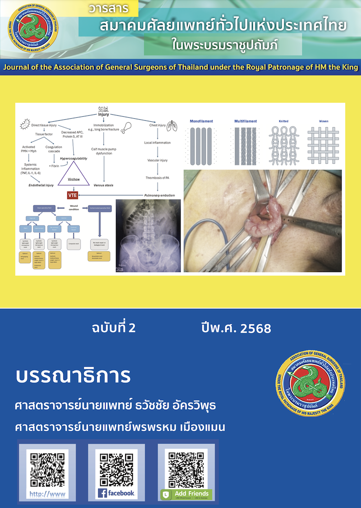
Background: Diabetic foot ulcers are a common and challenging complication in patients with diabetes mellitus, often complicated by infection and poor wound healing. Effective management requires adequate infection control, wound bed preparation, and optimized healing environment.
Case Presentation: A 67-year-old female with newly diagnosed type 2 diabetes mellitus presented with a non-healing plantar foot ulcer persisting for three weeks following hot water immersion. The wound was complicated by systemic infection and exposed plantar fascia. Surgical debridement was performed to remove necrotic tissue and reduce bacterial load. Wound cleansing with Prontosan® solution was employed to prevent biofilm formation. Periwound hyperkeratosis was trimmed, and dressings including Askina® Dressil and Askina® Heel were applied for moisture management and pressure offloading. Vascular assessments (ABI, TBI, TCOM) confirmed adequate perfusion. A biological skin substitute was subsequently used to promote tissue regeneration and wound closure.
Results: Infection was effectively controlled, and granulation tissue formation was observed over the previously exposed fascia. Minimal biofilm was managed with Prontosan® irrigation. The wound depth decreased rapidly after application of the skin substitute, with accelerated healing noted without need for complex reconstructive surgery.
Conclusion: Comprehensive wound care—including debridement, infection control, and the use of Prontosan® for biofilm management—plays a key role in treating complicated diabetic foot ulcers. Prontosan® may accelerate healing by reducing microbial burden, while advanced techniques help improve outcomes and limit invasive procedures.
References
Spampinato SF, Caruso GI, De Pasquale R, Sortino MA, Merlo S. The Treatment of Impaired Wound Healing in Diabetes: Looking among Old Drugs. Pharmaceuticals (Basel, Switzerland). 2020;13(4):60.
Akkus G, Sert M. Diabetic foot ulcers: A devastating complication of diabetes mellitus continues non-stop in spite of new medical treatment modalities. World J diabetes. 2022;13(12):1106-21.
Wang X, Yuan CX, Xu B, Yu Z. Diabetic foot ulcers: Classification, risk factors and management. World J diabetes. 2022;13(12):1049-65.
Cowan LJ, Stechmiller JK, Phillips P, Yang Q, Schultz G. Chronic Wounds, Biofilms and Use of Medicinal Larvae. Ulcers. 2013;2013(1):487024.
Senevirathne S, Ekanayake G, Samarathunge D, Basnayke O. The Use of Polyhexanide and Betaine Combined Preparation in Adult Burn Care in Sri Lanka. Cureus. 2024;16(8):e67274.
Atkin L, Stephenson J, Cooper DM. Wound bed preparation: a case series using polyhexanide and betaine solution and gel-a UK perspective. J Wound Care. 2020;29(7):380-6.
Daidone C, Salim N, Smith L, Raza A. The Role of Fish Skin Xenografts in Healing Complex Wounds: A Brief Case Report. Cureus. 2024;16(3):e56156.
Fiakos G, Kuang Z, Lo E. Improved skin regeneration with acellular fish skin grafts. Eng Regen. 2020;1:95-101.
Xiang J, Wang S, He Y, Xu L, Zhang S, Tang Z. Reasonable Glycemic Control Would Help Wound Healing During the Treatment of Diabetic Foot Ulcers. Diabetes Ther. 2019;10(1):95-105.
Watson F, Chen R, Saint Bezard J, Percival S. Comparison of antimicrobial efficacy and therapeutic index properties for common wound cleansing solutions, focusing on solutions containing PHMB. GMS Hyg Infect Control. 2024;19:Doc73.
Dueppers P, Bozalka R, Kopp R, Menges AL, Reutersberg B, Schrimpf C, et al. The Use of Intact Fish Skin Grafts in the Treatment of Necrotizing Fasciitis of the Leg: Early Clinical Experience and Literature Review on Indications for Intact Fish Skin Grafts. J Clin Med. 2023;12(18):6001.