
Optimization of radiation dose and image quality in abdominal radiography using digital mobile x-ray system
Keywords:
optimization, digital mobile x-ray system, abdominal radiography, entrance surface air kerma (ESAK)Abstract
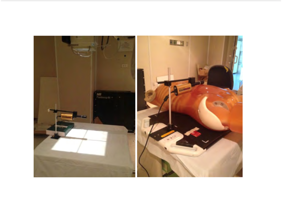
The purpose of this study was to optimize the radiation dose and image quality for abdominal radiography using digital mobile x-ray system in anthropomorphic phantom at King Chulalongkorn Memorial Hospital (KCMH). Digital mobile x-ray system model OptimaXR220amx, and the Kyoto Kagaku phantom model PBU-60 with 21 cm of abdominal thickness were used. The exposure parameters were set between 70-90 kVp, and 3.2, 6.3, 12.5, 25, and 32 mAs. Source to image receptor distance (SID) was set at 100 cm. The entrance surface air kerma (ESAK) in experimental and routine parameters were measured using ionization chamber (IC) dosimeter manufactured Radcal model ACCU-Gold. The qualitative image quality criteria based on IAEA protocol and qualitative were scored by three observers. The signal-to-noise ratio (SNR) was measured by placing three regions of interest at liver, transvers process at 4th lumbar spine, and pelvis respectively. The contrast-to-noise ratio (CNR) was evaluated on three areas at liver, left kidney and transverse process at 4th lumbar spine, right kidney and pelvis. We found that the optimal parameter for 21 cm thickness of abdomen at 100 cm SID was 80 kVp, 6.3 mAs with the exposure index of 381. The average image quality and qualitative noise scoring from three observers were 5.67 and 1, respectively. The average SNR in 1st, 2nd, and 3rd ROIs were 39.56, 61.55, and 18.24, respectively, and the average of CNR at 1st, 2nd, and 3rd areas were 5.97, 7.27, and 1.50, respectively. The ESAK obtained from optimal parameter was lower than 77% compared to the routine clinical parameter. The optimal exposure parameters in this study, however, can maintain the image quality with acceptable diagnosis for portable abdominal radiography.
Downloads
References
Seibert JA, Morin RL. The standardized exposure index for digital radiography: an opportunity for optimization of radiation dose to the pediatric population. Pediatr Radiol 2011;41:573-581.
Uffmann M, Schaefer-Prokop C. Digital radiography: the balance between image quality and required radiation dose. Eur J Radiol 2009;72:202-208.
Moore QT, Don S, Goske MJ, Strauss KJ, Cohen M, Herrmann T, et al. Image gently: using exposure indicators to improve pediatric digital radiography. Radiol Technol 2012;84:93-99.
Sangdao P. Optimization of radiation dose and image quality in chest radiography using digital mobile x-ray system at King Chulalongkorn Memorial Hospital [Thesis]. Bangkok: Chulalongkorn University; 2014.
International Atomic Energy Agency. Dosimetry in diagnostic radiology: An international code of practice. Technical report series (TRS) 457. Vienna, Austria 2007.
International Atomic Energy Agency. Radiation protection in digital radiology. Examples of good and bad image quality images with quality evaluation (projection radiography).
Aldrich JE, Duran E, Dunlop P, Mayo JR. Optimization of dose and image quality for computed radiography and digital radiography. J Digit Imaging 2006;19:126-131.
GE Healthcare. DEI (Detector Exposure Indicator). Optima XR200amx X-Ray System with Digital Upgrade Operator Manual 2012:14-41-14-44.
Muhogora WE, Ahmed NA, Almosabihi A, Alsuwaidi JS, Beganovic A, Ciraj-Bjelac O, et al. Patient doses in radiographic examinations in 12 countries in Asia, Africa, and Eastern Europe: initial results from IAEA projects. AJR Am J Roentgenol 2008;190:1453-61.
Shepard SJ, Wang J, Flynn M, Gingold E, Goldman L, Krugh K, et al. An exposure indicator for digital radiography: AAPM Task Group 116 (executive summary). Med Phys 2009;36:2898-914.
Asada Y, Suzuki S, Minami K, Shirakawa S, Kobayashi M. Survey of patient exposure from general radiography and mammography in Japan in 2014. J Radiol Prot 2016;36:N8-N18.
Masoud AO, Muhogora WE, Msaki PK. Assessment of patient dose and optimization levels in chest and abdomen CR examinations at referral hospitals in Tanzania. J Appl Clin Med Phys 2015;16:5614.
Downloads
Published
How to Cite
Issue
Section
License
บทความที่ได้รับการตีพิมพ์เป็นลิขสิทธิ์ของสมาคมรังสีเทคนิคแห่งประเทศไทย (The Thai Society of Radiological Technologists)
ข้อความที่ปรากฏในบทความแต่ละเรื่องในวารสารวิชาการเล่มนี้เป็นความคิดเห็นส่วนตัวของผู้เขียนแต่ละท่านไม่เกี่ยวข้องกับสมาคมรังสีเทคนิคแห่งประเทศไทยและบุคคลากรท่านอื่น ๆในสมาคม ฯ แต่อย่างใด ความรับผิดชอบองค์ประกอบทั้งหมดของบทความแต่ละเรื่องเป็นของผู้เขียนแต่ละท่าน หากมีความผิดพลาดใดๆ ผู้เขียนแต่ละท่านจะรับผิดชอบบทความของตนเองแต่ผู้เดียว


