
Comparison of computed tomography dose index measuring by two detector types of computed tomography simulator
Keywords:
Computed tomography simulator, CTDIvol, Ionization chamber, Solid state detectorAbstract
Introduction: In general, the computed tomography screen displays the volume CT dose index (CTDIvol) received from a computed tomography by following the technique used. Verifying the display values is important before using them. This can be done by measuring the amount of radiation that is presented on the screen. However, there are 2 types of radiation detectors i.e., gas-filled and solid media types. These 2 detectors provide different methods of measurement and calculation. Objective: The purpose of this study was to compare the difference in CTDIvol values obtained from both detectors. Materials and methods: The CTDIvol was measured by using the ionization chamber and semiconductor detector in a phantom computed tomography 16 slices for Optima 580 simulation of GE Health Care Thailand. The CT setting parameters were based on the brain scan technique and the abdominal cavity using in the treatment planning simulation. The values obtained from the displayed on the screen of CT and measured from the detectors were compared and calculated as the percentage difference in CTDIvol. Results: Solid-state detector measured CTDIvol radiation less than conventional gas-filled detector. The different percentages were 10.20 and 5.02 for brain and abdominal cavity examination respectively. Conclusion: Radiation doses from computed tomography simulator measured by solid-state detector are less than those measured by standard gaseous radiation detector.
Downloads
References
American Association of Physicists in Medicine (AAPM) Report No.96 The Measurement, Reporting, and Management of Radiation Dose in CT. College Park: AAPM; 2008.
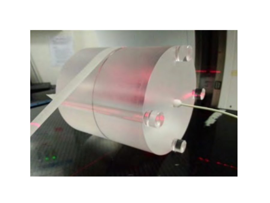
CIRS CT Dose Phantom. Chesterland: LACO, Inc; [updated 2017; cited 2017 May 15]. Available from: http://www.lacoonline.com/cgi-bin/lacoonline/00177.html.
International Atomic Energy Agency (IAEA) human health series No.19 Quality assurance programme for computed tomography diagnostic and therapy applications. Austria: IAEA; 2012.
National Council on Radiation Protection and Measurements. NCRP Report No. 99 Quality Assurance for Diagnostic Imaging. USA: NCRP; 1998.
Technical reports series No.457 Dosimetry in diagnostic radiology: an international code of practice. Austria: International Atomic Energy Agency (IAEA); 2007.
CT Dose Profiler user’s manual. Sweden: RTI ElectronicAB; 2015.
Grönlund E. Calibration & Clinical Measurements in Computed Tomography-An Evaluation of Different Dosimetric Methods: University of Gothamburk; 2013.
Downloads
Published
How to Cite
Issue
Section
License
บทความที่ได้รับการตีพิมพ์เป็นลิขสิทธิ์ของสมาคมรังสีเทคนิคแห่งประเทศไทย (The Thai Society of Radiological Technologists)
ข้อความที่ปรากฏในบทความแต่ละเรื่องในวารสารวิชาการเล่มนี้เป็นความคิดเห็นส่วนตัวของผู้เขียนแต่ละท่านไม่เกี่ยวข้องกับสมาคมรังสีเทคนิคแห่งประเทศไทยและบุคคลากรท่านอื่น ๆในสมาคม ฯ แต่อย่างใด ความรับผิดชอบองค์ประกอบทั้งหมดของบทความแต่ละเรื่องเป็นของผู้เขียนแต่ละท่าน หากมีความผิดพลาดใดๆ ผู้เขียนแต่ละท่านจะรับผิดชอบบทความของตนเองแต่ผู้เดียว


