
Development of breast lesion detection program
Keywords:
Breast mass, Breast lesion prediction, Mammography, MicrocalcificationAbstract
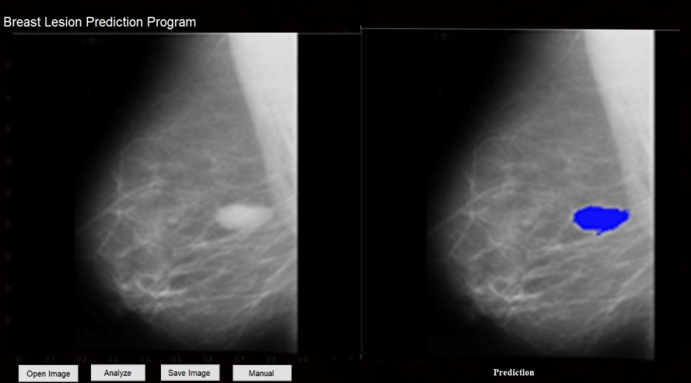
Since the diversity of lesions represented in the breast and lesions that are shown in mammography were not clear in some cases and were similar to surrounding normal tissue which will cause to missing of diagnosis and specifying lesion localization, which resulted in difficulty of analysis in mammography. This study aimed to develop a program that use of Fuzzy C-means combined with morphological techniques to predict breast lesion. By using the sample of 213 mammographic images in Mediolateral Oblique view (MLO) with lesions type (Mass, Microcalcification or Normal) from the online database (The Mini-MIAS), the mammographs were divided into 3 groups (40 mammographs for Mass lesion, 17 Mammographs for Microcalcification lesion and 156 mammographic images for Normal which is not shown for lesion). The program was analyzed the pictures by using an algorithm that was set for specific lesion localization and predict the breast lesion represents in Mass, Microcalcification and Normal, and measured the sensitivity, specificity, and accuracy. The results show that sensitivity of mass and Microcalcification detection rate were 52.5% and 0%, respectively, specificity was 66.0% and accuracy was 63.2 and 59.5%, respectively.
Downloads
References
นพ.สาธิต ศรีมันทยามาศ. 5 ความเข้าใจผิด เกี่ยวกับมะเร็งเต้านม. [Internet]. [cited 2018 April 20]. Available from: https://mgronline.com/qol/detail/9590000100312.
การเอกซเรย์เต้านม (Mammogram). [Internet]. [cited 2018 April 20]. Available from:
http://iamradiographer.blogspot.com/2010/11/mammogram.html.
The mini-MIAS database of mammograms. [Internet]. [cited 2018 April 20]. Available from:
http://peipa.essex.ac.uk/info/mias.html
ใจภักดิ์ ติรเดชาฤทธิ์,วิรัญชนา ฉัตรจันทึกและอภิเชฐษ์ เวียนเป๊ะ. การแยกรอยโรคของเต้านมโดยใช้เทคนิคฟัซซี่ ซี-มีนส์ ในภาพเอกซเรย์เต้านม [วิทยานิพนธ์]. พิษณุโลก: ภาควิชารังสีเทคนิค คณะสหเวชศาสตร์ มหาวิทยาลัยนเรศวร; 2556.
นิตยา นาคเย, พกาวรรณ ไพรทอง และสุภาพร จันทะ. การศึกษาข้อมูลภาพเชิงปริมาณของภาพเอกซเรย์เต้านม [วิทยานิพนธ์]. พิษณุโลก: ภาควิชารังสีเทคนิค คณะสหเวชศาสตร์ มหาวิทยาลัยนเรศวร; 2556.
ฐิติพงศ์ แก้วเหล็ก นิตยา นาคเย พกาวรรณ ไพรทอง และ สุภาพร จันทะ. ผลการศึกษาข้อมูลเชิงสถิติของภาพเอกซเรย์เต้านม. ศรีนครินทร์เวชสาร. 2562 :34(1).
Downloads
Published
How to Cite
Issue
Section
License
บทความที่ได้รับการตีพิมพ์เป็นลิขสิทธิ์ของสมาคมรังสีเทคนิคแห่งประเทศไทย (The Thai Society of Radiological Technologists)
ข้อความที่ปรากฏในบทความแต่ละเรื่องในวารสารวิชาการเล่มนี้เป็นความคิดเห็นส่วนตัวของผู้เขียนแต่ละท่านไม่เกี่ยวข้องกับสมาคมรังสีเทคนิคแห่งประเทศไทยและบุคคลากรท่านอื่น ๆในสมาคม ฯ แต่อย่างใด ความรับผิดชอบองค์ประกอบทั้งหมดของบทความแต่ละเรื่องเป็นของผู้เขียนแต่ละท่าน หากมีความผิดพลาดใดๆ ผู้เขียนแต่ละท่านจะรับผิดชอบบทความของตนเองแต่ผู้เดียว


