
The study of kilovoltage peak selection for chest x-ray examination in Samutprakarn province
Keywords:
Peak tube kilovoltage, penetrating power, chest x-rays, exposure techniqueAbstract
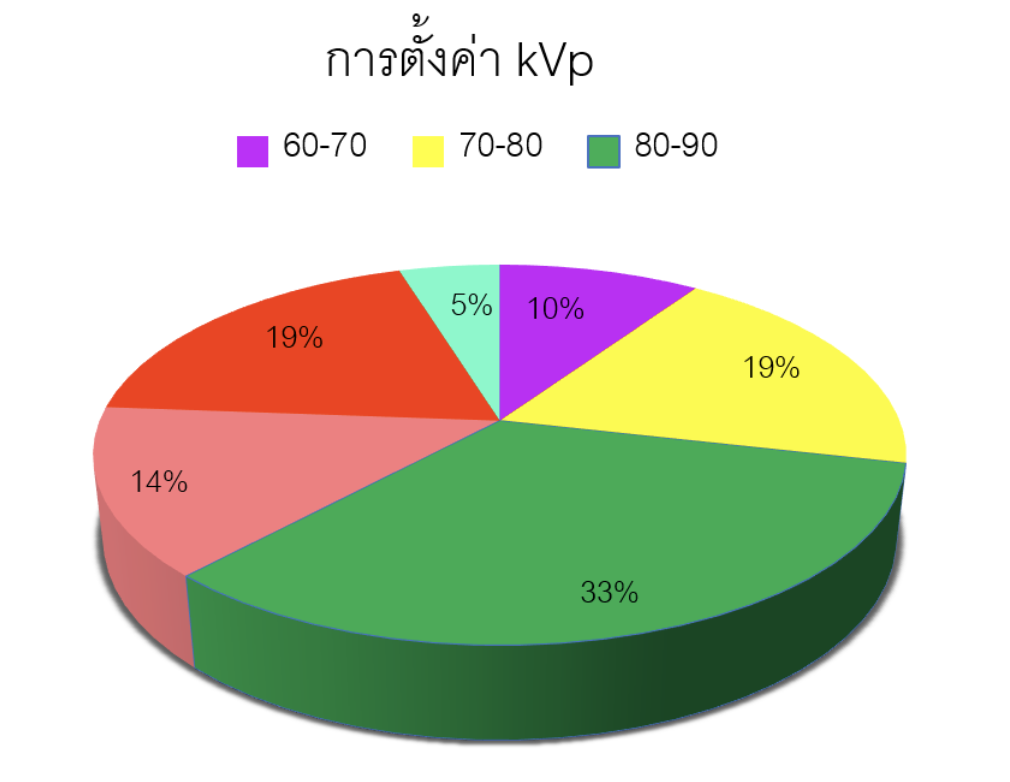
Peak kilovoltage (kVp) is one of the exposure techniques used to control an image quality of chest x-ray (CXR) and an exposure to patient. In the past, the kVp was strictly selected based on screen-film characteristics to maintain a good radiographic quality. Digital imaging system can provide wider range of kVp selection while maintaining adjustable display image. This would encourage new technique such as high kVp CXR which provides better lung parenchyma and mediastinum seen. Samutprakarn province is an important industrial area that usually require CXR for health screening of the staff. We have noticed that kVp for CXR of each hospital did not similar event in nearby area. This study aimed to survey the kVp for CXR and factors related to these kVp selection from 21 hospitals in Samutprakarn province using the questionnaire. The results showed the mode kVp was 85 and the average kVp was 87.85 ± 13.83. The highest satisfactions score was 4.19 ± 0.68 (Image detail). The lowest satisfactions score was 2.42 ± 1.16 (screen-films technique adopting). There was significant correlation between body part thickness and penetrating power (r = 0.615, p < 0.05) and image detail. There was significant correlation between detectability of radiographic pathology (r = 0.612, p < 0.05). The high tube voltage range between 110-110 kVp was selected by the radiologist while the low range between 70 – 90 kVp was selected among radiographers. The mode kVp (85) in this study was not high kVp technique. The wide kVp range of 70 – 120 had shown the variation of kVp selection for CXR in Samutprakarn hospital. Further study should include patient exposure and clinical information.
Downloads
References
(1) สำนักงานจังหวัดสมุทรปราการ. ข้อมูลทั่วไปของจังหวัดสมุทรปราการ [อินเตอร์เน็ต]. มิถุนายน. 2554 [เข้าถึงเมื่อวันที่ 21 มิถุนายน 2562]. เข้าถึงได้จาก: http://www.samutprakan.go.th/newweb/index.php?option=com_content&view=article&id=67:2011-11-29-15-59-12&catid=14:2011-11-29-15-17-14&Itemid=12
(2) สำนักงานแรงงาน จังหวัดสมุทรปราการ. สภาพเศรษฐกิจจังหวัดสมุทรปราการ [อินเตอร์เน็ต].2560 [เข้าถึงเมื่อวันที่ 21 มิถุนายน 2562].
เข้าถึงได้จาก https://samutprakan.mol.go.th/sites/samutprakan.mol.go.th/files/5.enuuehaaaitrmaas1.pdf
(3) Anouk M Speets, Yolanda van der Graaf, Arno W Hoes, Sandra Kalmijn, Alfred PE Sachs, Matthieu JCM Rutten, et al. Chest radiography in general practice: indications, diagnostic yield and consequences for patient management. British Journal of General Practice, August 2006
(4) Walter Huda, R. Brad Abrahams. Radiographic Techniques, Contrast, and Noise in X-Ray Imaging. AJR:204, February 2015
(5) Honey ID, Mackenzie A, Evans DS. Investigation of optimum energies for chest imaging using film-screen and computed radiography. Br J Radiol 2005;78(929):422-7. doi: 10.1259/bjr/32912696. PMID: 15845936.
(6) Lorusso JR, Fitzgeorge L, Lorusso D, Lorusso E. Examining Practitioners' Assessments of Perceived Aesthetic and Diagnostic Quality of High kVp-Low mAs Pelvis, Chest, Skull, and Hand Phantom Radiographs. J Med Imaging Radiat Sci 2015;46(2):162-173.
doi: 10.1016/j.jmir.2015.01.109. PMID: 31052090.
(7) Xiaoming Zheng, Myeongsoo Kim, Sook Yang, "Optimal kVp in chest computed radiography using visual grading scores: a comparison between visual grading characteristics and ordinal regression analysis," Proc. SPIE 9783, Medical Imaging 2016: https://doi.org/10.1117/12.2217414
(8) เพ็ญศิลา สุภาพ. การศึกษาเทคนิคการตั้งค่าปริมาณรังสีที่เหมาะสมและปริมาณรังสีที่ผู้ป่วยได้รับของการถ่ายภาพรังสีทรวงอกในผู้ป่วยสภาพปกติโรงพยาบาลกันทรลักษณ์ จังหวัดศรีษะเกษ; 2556.
เข้าถึงได้จาก: http://203.157.165.4/ssko_presents/file_presents/3330200120494-8-1097.doc
(9) ลัดดา เย็นศรี. การศึกษาเปรียบเทียบปริมาณรังสีที่ผู้ป่วยได้รับจากการถ่ายภาพรังสีทรวงอกด้วยระบบ DR และ CR. วารสารเครือข่ายวิทยาลัยพยาบาลและการสาธารณสุข ภาคใต้ 2559;3(1):1
(10) Zoe Brady, Heather Scoullar, Ben Grinsted, Kyle Ewert, Helen Kavnoudias, Alexander Jarema, et al. Technique, radiation safety and image quality for chest X‑ray imaging through glass and in mobile settings during the COVID‑19 pandemic. Physical and Engineering Sciences in Medicine (2020) 43:765–779 https://doi.org/10.1007/s13246-020-00899-8
Downloads
Published
How to Cite
Issue
Section
License
บทความที่ได้รับการตีพิมพ์เป็นลิขสิทธิ์ของสมาคมรังสีเทคนิคแห่งประเทศไทย (The Thai Society of Radiological Technologists)
ข้อความที่ปรากฏในบทความแต่ละเรื่องในวารสารวิชาการเล่มนี้เป็นความคิดเห็นส่วนตัวของผู้เขียนแต่ละท่านไม่เกี่ยวข้องกับสมาคมรังสีเทคนิคแห่งประเทศไทยและบุคคลากรท่านอื่น ๆในสมาคม ฯ แต่อย่างใด ความรับผิดชอบองค์ประกอบทั้งหมดของบทความแต่ละเรื่องเป็นของผู้เขียนแต่ละท่าน หากมีความผิดพลาดใดๆ ผู้เขียนแต่ละท่านจะรับผิดชอบบทความของตนเองแต่ผู้เดียว


