
Comparison between conventional pre-contrast and virtual non-contrast images from IQon spectral CT
Keywords:
Spectral computed tomography, Virtual non-contrast, True non-contrast, CT numberAbstract
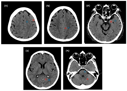
Dual-layer CT is a dual-energy CT is technology that uses the dual layer detector to generate two different tube voltage image. The image raw data can be reconstructed many image types, such as virtual mono-energetic image, virtual non-contrast image, etc. This study aimed to evaluate the difference in the CT number between the pre-contrast CT image or True non-contrast (TNC) and Virtual non-contrast (VNC) of the brain. The retrospective data of 35 patients who underwent CT brain without and with contrast media were collected from Philips IQon Spectral CT at Advanced Diagnostic Imaging Center. CT number values of white matter (WM) and gray matter (GM) of TNC and VNC CT images were measured at both left and right hemispheres of the frontal lobe, parietal lobe, temporal lobe, occipital lobe, and pons (WM) & cerebellum (GM) and compared with T-test (two-tailed) with the level of confidence p-value 0.05. The patient’s average and standard deviation were 65.6±7.8 years, and CTDIvol ranged from 45.5 to 58.3 mGy. The study found that the average CT numbers at all measured positions in TNC and VNC images were 27.1±1.3 and 27.1±1.8 HU for white matter, and 34.0±1.6 and 28.9±2.6 HU for gray matter, respectively. CT numbers of white matter was no significant difference (p=0.05) and gray matter was differ significant (<0.05). Conclusions, the image contrast of white and gray matter in VNC images is inferior to TNC images. It is concluded that using the VNC image of the brain instead of the TNC image had to be particularly cautions, especially in the case of white and gray matter diagnoses. However, it may be used in the followed-up case to reduce the patient’s dose.
Downloads
References
Johnson T. R. C.. Dual-Energy CT: General Principles. AJR Am J Roentgenol 2012;199(5, Suppl):S3–S8.
Mangesius S., et al. Dual-energy computed tomography in acute ischemic stroke: state-of-the-art. Eur. Radiol. 2021; 31:4138–4147.
McCollough CH, Leng S, Yu L, Fletcher JG. Dual-and multi-energy CT: principles, technical approaches, and clinical applications. Radiology 2015;276(3):637-53.
Postma AA, Das M, Stadler AA, Wildberger JE. Dual-energy CT: what the neuroradiologist should know. Curr Radiol Rep 2015;3(5):16.
Naruto N, Itoh T, Noguchi K. Dual energy computed tomography for the head. Jpn J Radiol 2018;36(2):69-80.
Angelo TD., et al. Dual energy computed tomography virtual monoenergetic imaging: technique and clinical applications. Br J Radiol 2019; 92:20180546
Albrecht MH., et al. Review of Clinical Applications for Virtual Monoenergetic Dual-Energy CT. Radiology 2019; 293:260–271.
Weinstein MA, Duchesneau PM, MacIntyre WJ. White and gray matter of the brain differentiated by computed tomography. Radiology 1977;122(3):699-702.
Jiang XY, et al. Evaluation of Virtual Noncontrast images obtained from dual-energy CTA for diagnosing subarachnoid hemorrahge. AJNR Am J Neuroradiol May 2015; 36:855–60.
Niehoff JH., Woeltjen MM., Laukamp KR. And Borggrefe J. Virtual non-contrast versus true non-contrast computed tomography: initial experience with photon counting scanner approved for clinical use. Diagnostics 2021, 11, 2377.
Kanal KM, Butler PF, Sengupta D, Bhargavan-Chatfield M, Coombs LP, Morin RL. US diagnostic reference levels and achievable doses for 10 adult CT examinations. Radiology 2017;284(1):120-33.
Dance D, Christofides S, Maidment A, McLean I, Ng K. Diagnostic radiology physics: A Handbook for Teachers and Students. IAEA Vienna, 2014: 260.
Downloads
Published
How to Cite
Issue
Section
License
Copyright (c) 2022 The Thai Society of Radiological Technologists

This work is licensed under a Creative Commons Attribution-NonCommercial-NoDerivatives 4.0 International License.
บทความที่ได้รับการตีพิมพ์เป็นลิขสิทธิ์ของสมาคมรังสีเทคนิคแห่งประเทศไทย (The Thai Society of Radiological Technologists)
ข้อความที่ปรากฏในบทความแต่ละเรื่องในวารสารวิชาการเล่มนี้เป็นความคิดเห็นส่วนตัวของผู้เขียนแต่ละท่านไม่เกี่ยวข้องกับสมาคมรังสีเทคนิคแห่งประเทศไทยและบุคคลากรท่านอื่น ๆในสมาคม ฯ แต่อย่างใด ความรับผิดชอบองค์ประกอบทั้งหมดของบทความแต่ละเรื่องเป็นของผู้เขียนแต่ละท่าน หากมีความผิดพลาดใดๆ ผู้เขียนแต่ละท่านจะรับผิดชอบบทความของตนเองแต่ผู้เดียว


