
The comparative study of left ventricular ejection fraction (LVEF) between gated blood pool tomography and multiple gated acquisition (MUGA) scan
Keywords:
Left ventricular ejection fraction, Gated blood pool tomography, Multiple gated acquisitionAbstract
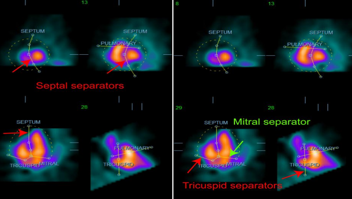
Background: MUGA scan is the nuclear medicine technique used to evaluate left ventricular ejection fraction of the heart. It had high reproducibility in each examination. In cases which MUGA is failed or incorrect result due to tumor obscured the heart, GBPS (gated blood-pool SPECT) is an alternative method. GBPS is not routine practiced at King Chulalongkorn Memorial Hospital (KCMH) service. It is according to capability of scanner and software package for processing. Now, KCMH has the scanner that has this capability but no experience to perform processing and the reliability of results. This study will help technologist to make confidence in processing and nuclear medicine physician to interpret result. Methods: Patients with request for pre- chemotherapy %LVEF base line and volunteer, who did not on drug which affect red blood cell labeling with technetium pertechnetate, were included in this study. The method of RBC (red blood cell) labeling was modified in vivo technique. Perform MUGA acquisition in LAO (left anterior oblique) view with best septal seen. Preset 1000 heart beats with 64 x 64 matrix, zoom 1.45 with LEUR (low energy ultra-high resolution) collimator on Siemens Symbia T camera and followed by GBPS acquisition with 64 x 64 matrix, zoom 1.45, 64 views over 180 degree rotation arc, duration 50 sec/view, perpendicular configuration detector and start from RAO (right anterior oblique)45 degree. Perform reconstruction and processing for nuclear medicine physician interpretation. Results: Total study was 55 cases (17 males and 38 females) with the age (mean±SD) was 44.38±10.54 years. The relationship between MUGA and GBPS was good correlation with R =0.84. Nuclear medicine physician interpreted 54 cases were within normal limits and one case was abnormal %LVEF. The mean normal value of %LVEF by MUGA was 65.67±4.43 and GBPS was 77.33±7.0. Conclusion: The Patient whom fail to perform MUGA processing can used GBPS for %LVEF evaluation. %LVEF by GBPS was reproducible for automated processing and correlated well with MUGA with R =0.84. %LVEF by GBPS was higher than MUGA due to left atrium counts was included in MUGA calculation.
Downloads
References
Dutta T, Spevack DM, Aronow WS. The left ventricular ejection fraction: new insights into an old parameter. Hospital practice. 2019;47(5):221-230.
Pelletier-Galarneau M, Finnerty V, Tan S, Authier A, Eng B, Gregoirw J, et al. Assessment of left ventricular ejection fraction with cardiofocal collimators: Comparison between IQ-SPECT, planar equilibrium radionuclide angiography, and cardiac magnetic resonance. J Nucl Cardiol. 2019;26:1857-1864.
Royen NV, Jaffe CC, Krumholz HM, Johnson KM, Lynch PJ, Atkinson P, et al. Comparison and reproducibility of visual echocardiographic and quantitative radionuclide left ventricular ejection fractions. Am J Cardiol. 1996;77:843-850.
Mitra D, Basu S. Equilibrium radionuclide angiocardiography: Its usefulness in current practice and potential future applications. World J Radiol. 2012;4(10):421-430.
Perez IE, Alam ST, Hernandez GA, Sancassani R. Cancer Therapy Related Cardiac Dysfunction: An Overview for the Clinician. Clin Med Insights Cardiol. 2019;13:1-11.
Kolla BC, Roy SS, Duval S, Weisdorf D, Valeti U, Blaes A. Cardiac Imaging Methods for Chemotherapy-related Cardiotoxicity Screening and Related Radiation Exposure: Current Practice and Trends. Anticancer Res. 2017;37:2445-2449.
Yang SN, Sun SS, Zhang G, Chou KT, Lo SW, Chiou YR, et al. Left ventricular ejection fraction estimation using mutual information on technetium-99m multiple-gated SPECT scans. Biomed Eng Online. 2015;14:119:1-10.
Eeckhoudt SV, Ziekenhuis B, Schelfhout RV, Rijnstate, Arnhem. Equilibrium Radionuclide Angiography/Multigated acquisition. [Internet] Available from: https://richtlijnendatabase.nl/gerelateerde_documenten/f/17261/Equilibrium%20Radionuclide%20Angiography.pdf
Groch MW, Depuey EG, Belzberg AC, Erwin WD, Kamran M, Barnett CA, et al. Planar imaging versus gated blood-pool SPECT for the assessment of ventricular performance. A multicenter study. J Nucl Med. 2001;42:1773-1779.
Bartlett ML, Srinivassan G, Baker WC, Kitsiou AN, Dilsizian V, Bacharach SL. Left ventricular ejection fraction: comparison of results from planar and SPECT gated blood pool studies. J Nucl Med. 1996;37:1795-1799.
Cedars-Sinai Medical Center. QBS quantitative blood pool SPECT reference manual version 2012.1: October 2011, 8700 Beverly Blvd., CA, USA.
Downloads
Published
How to Cite
Issue
Section
License
Copyright (c) 2023 The Thai Society of Radiological Technologists

This work is licensed under a Creative Commons Attribution-NonCommercial-NoDerivatives 4.0 International License.
บทความที่ได้รับการตีพิมพ์เป็นลิขสิทธิ์ของสมาคมรังสีเทคนิคแห่งประเทศไทย (The Thai Society of Radiological Technologists)
ข้อความที่ปรากฏในบทความแต่ละเรื่องในวารสารวิชาการเล่มนี้เป็นความคิดเห็นส่วนตัวของผู้เขียนแต่ละท่านไม่เกี่ยวข้องกับสมาคมรังสีเทคนิคแห่งประเทศไทยและบุคคลากรท่านอื่น ๆในสมาคม ฯ แต่อย่างใด ความรับผิดชอบองค์ประกอบทั้งหมดของบทความแต่ละเรื่องเป็นของผู้เขียนแต่ละท่าน หากมีความผิดพลาดใดๆ ผู้เขียนแต่ละท่านจะรับผิดชอบบทความของตนเองแต่ผู้เดียว


