
The determination of the radiation dose and image quality for abdominal radiography using a digital X-ray machine without an automatic exposure control system at King Chulalongkorn Memorial Hospital
Keywords:
Automatic exposure control, Anthropomorphic phantom, Entrance surface air kerma, Kerma area productAbstract
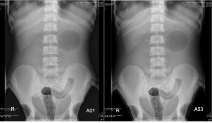
Background: Radiographic procedures with automatic exposure control (AEC) system are the exposure technique to obtain the optimal diagnostic image quality. However, there is a higher dose under AEC compared with a lack of AEC. Objective: The aim of this study was to determine the radiation dose and image quality for abdominal digital radiography of human-like phantom using a routine clinical protocol with AEC and a modified protocol without AEC at King Chulalongkorn Memorial Hospital. Methods: A digital X-ray system (Digital Diagnost, Philips) with a CsI:Ti image receptor and the human-like phantom (PBU-60, Kyoto Kagaku) were used. The abdominal anteroposterior (AP) projections were performed with and without AEC systems. The entrance surface air kerma (ESAK) was measured on the phantom surface using optically stimulated luminescence dosimeters (OSLDs), while the kerma area product (KAP) was simultaneously recorded. The qualitative image quality was scored by two experienced radiologists. Results: We found that the ESAK and KAP values trended to the similar direction. The ESAK obtained from the modified parameter without AEC (81 kVp and 3.2 mAs) was lower by 90% compared to the routine clinical parameter with AEC (85 kVp and 32 mAs), while the optimal image quality was maintained in the acceptable level for diagnostic abdominal radiography. Conclusion: Applying the exposure parameter without AEC for abdominal digital radiography reduces the radiation while the image quality is optimal for diagnosis.
Downloads
References
International Atomic Energy Agency. Avoidance of unnecessary dose to patients while transitioning from analogue to digital radiography. Vienna, IAEA-TECDOC-1667. 2011.
Grewal RK, Young N, Colins L, Karunnaratne N, Sabharwal N. Digital chest radiography image quality assessment with dose reduction. Australas Phys Eng Sci Med. 2012; 35: 71-80.
Choi SS, Lim CH, Jeoung SH. Automatic exposure control in chest radiography. J Econ Lit. 2019; 19(1): DOI: 10.5958/0974-1283.2019.00125.7.
Kim H, Park M, Park S, Jeong H, Kim J, Kim Y. Estimation of absorbed organ doses and effective dose based on body mass index in digital radiography. Radiat Prot Dosimetry. 2013; 153: 92-9.
Suwan-o-pas S, Suwanpradit P, Arjhansiri K, Khamwan K. Optimization of radiation dose and image quality in abdominal radiography using digital mobile x-ray system. Thai J Rad Tech. 2018; 43(1): 13-20.
Kyoto Kagaku, Available from: https://www/kyotokagaku.com/en/products_data/ph-2b/
Microstar user guide version 5.0. Laudauer; 2015.
European commission. European guidelines on quality criteria for diagnostic radiographic images. Luxembourg, EUR 16260 EN. 1996.
Hallgren KA. Computing inter-rater reliability for observational data: an overview and tutorial. Tutor Quant Methods Psychol. 2012; 8(1): 23-34.
Landis JR, Koch GG. The measurement of observe agreement for categorical data. Biometrics. 1977; 33(1): 159-74.
Jang JS, Koo HJ, Yang HJ, Park JH, Cho YC, Do KH. Effective dose in abdominal digital radiography: patient factors. J Korean Soc Radiol. 2017; 77(2): 89-96.
Jang JS, Yang HJ, Koo HJ, Kim SH, Park CR, Yoon SH, Shin SY, Do KH. Image quality assessment with dose reduction using high kVp and additional filtration for abdominal digital radiography. Phys Med. 2018; 50: 46-51.
Almaziad GA, Shahrani AA, Singh OG, Ayaz AA, Abudiab FI, Baligun SO. Modification of the projection from anterior-posterior to posterior-anterior for abdominal radiographic examinations. Biosc Biotech Res Comm. 2021; 14(5): 258-64.
Downloads
Published
How to Cite
Issue
Section
License
Copyright (c) 2023 The Thai Society of Radiological Technologists

This work is licensed under a Creative Commons Attribution-NonCommercial-NoDerivatives 4.0 International License.
บทความที่ได้รับการตีพิมพ์เป็นลิขสิทธิ์ของสมาคมรังสีเทคนิคแห่งประเทศไทย (The Thai Society of Radiological Technologists)
ข้อความที่ปรากฏในบทความแต่ละเรื่องในวารสารวิชาการเล่มนี้เป็นความคิดเห็นส่วนตัวของผู้เขียนแต่ละท่านไม่เกี่ยวข้องกับสมาคมรังสีเทคนิคแห่งประเทศไทยและบุคคลากรท่านอื่น ๆในสมาคม ฯ แต่อย่างใด ความรับผิดชอบองค์ประกอบทั้งหมดของบทความแต่ละเรื่องเป็นของผู้เขียนแต่ละท่าน หากมีความผิดพลาดใดๆ ผู้เขียนแต่ละท่านจะรับผิดชอบบทความของตนเองแต่ผู้เดียว


