
Quality control of chest radiograph procedure
Keywords:
Chest radiograph, Quality control, Long-length radiography, X-ray positioningAbstract
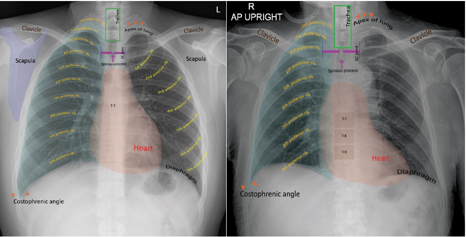
Chest radiography, also known as a chest x-ray. The images show the internal organs of the chest and neighboring structures. A chest radiograph which an X-ray beam enters behind the patient and exits the front of patient to an anterior image receptor, known as the Posteroanterior (PA) and X-ray beam passes from anterior the patient to the image receptor behind, known as the Anteroposterior (AP), is the most common radiological investigation for diagnosing chest abnormalities. As a guideline to reduce the repetitive rate of x-rays, radiological technologist should have knowledge and understanding for quality control of chest radiographs. Starting from patients’ factors that come in different conditions, which requires skill and experience in the work of radiological technologist to assess the appropriate positioning for each case. Somes requires a method of positioning that is different from the routine from the textbook including the factors from the radiological technologist that should be known to adjust the various parameters of the X-ray machine in order to provide the appropriate radiation dose from different positions in each patient. Additionally, the process of evaluating whether the obtained photographs are sufficient quality for the radiologist to report the accurately results. The factors to control the quality of chest radiographs should begin from the process of patient preparation, positioning, X-ray machine parameters setting and post image processing from the software of the x-ray machine to be accurately. If the radiological technologist does not have accurate knowledge of chest radiographs, this may cause in the radiologist receive improper image, resulting in inaccurate or unclear report affecting recipients to lose the opportunity to receive the correct diagnosis. Therefore, it is necessary to have standards to have quality control of chest radiographs before sending the images to the PACs system for radiologist’s report.
Downloads
References
Murphy A. Chest (PA view) [Internet]. 2023 [cited 2022 January 18]. Available from: https://radiopaedia.org/articles/chest-pa-view-1?lang=us
วัชรากร ปานเนียม, พงศ์พล ลิมป์พรวิเชียร, นัที อินา, จงวัฒน์ ชีวกุล, เสรี ศักดิ์จิรพาพงษ์. การศึกษาปัจจัยและแนวทางลดอัตราการถ่ายภาพเอกซเรย์ซ้ำในระบบเอกซเรย์ดิจิตอลของการถ่ายภาพเอกซเรย์ทรวงอกและเข่าท่ายืนด้านข้าง ณ โรงพยาบาลสงขลานครินทร์. วารสารรังสีเทคนิค. 2562;44(1):45-52.
กลุ่มงานรังสีวิทยา โรงพยาบาลลำพูน.การออกแบบและการสร้างอุปกรณ์ยึดจับตลับฟิล์มให้ผู้ป่วยที่นอนมาบนเปลนอนที่มีอาการสั่นไหวยึดเกาะเพื่อถ่ายภาพรังสีทรวงอกท่านั่ง [อินเทอร์เน็ต]. ลำพูน: กลุ่มงานรังสีวิทยา โรงพยาบาลลำพูน; 2557 [เข้าถึงเมื่อ 20 พ.ย. 2565]. เข้าถึงได้จาก: http://203.157.203.2/archivesImages/2557/138errand/55116_67_o.pdf
สมหมาย กันทะเมืองลี้. การวิเคราะห์ภาพดิจิตอลทางรังสีที่ไม่สามารถนำไปวินิจฉัยโรคได้ (reject image) โรงพยาบาลนครปฐม. วารสารสุขภาพภาคประชาชน. 2560;12(4):28-35.
กรมวิทยาศาสตร์การแพทย์ กระทรวงสาธารณสุข. การควบคุมคุณภาพเครื่องเอกซเรย์วินิจฉัยทางการแพทย์ 2565. กรุงเทพฯ: บริษัท บียอนด์ พับลิสซิ่ง จำกัด; 2565.
Lim I. AP CHEST X-RAY (Supine or Semierect in Department or as Beside Portable) [Internet]. 2011 [cited 2020 January 22]. Available from: http://www.radtechonduty.com/2011/12/ap-chest.html
Murphy A. Chest (AP erect view) [Internet]. 2021 [cited 2022 January 11]. Available from: https://radiopaedia.org/articles/chest-ap-erect-view-1?lang=us
ศุภวรรณ จิวะพงศ์, บุณยานุช พัฒนาดิสัย, สมลักษณ์ จำรูญสาย. ปริมาณรังสีที่ผู้ป่วยได้รับจากการถ่าย ภาพรังสีทรวงอกที่งานรังสีเทคนิค ศูนย์การแพทย์กาญจนาภิเษก. เวชบันทึกศิริราช. 2566;16(1):10-7.
วรรณพร บุรีวงษ์. รังสีวิทยาระบบทางเดินหายใจ: การเลือกส่งตรวจและแปลผลภาพรังสีทรวงอก (Radiology of the chest: methods of investigation and plain film interpretation) [อินเตอร์เน็ต]. 2557. [เข้าถึงเมื่อ 28 ธ.ค. 2565]. เข้าถึงได้จาก: http://med.swu.ac.th/radiology/images/stories/Education/chest%20%20cvs%202014.pdf
Williams MB, Krupinski EA, Strauss KJ, Breeden WK, 3rd, Rzeszotarski MS, Applegate K, et al. Digital radiography image quality: image acquisition. Journal of the American College of Radiology: JACR. 2007;4(6):371-88.
Lim I. Chest: PA View [Internet]. 2014 [cited 2022 February 10]. Available from: http://www.radtechonduty.com/2011/12/pa-projection-chest.html
Murphy A. Chest (AP supine) [Internet]. 2023 [cited 2021 December 23]. Available from: https://radiopaedia.org/articles/chest-supine-view-1?lang=us
Murphy A. Ribs (PA view) [Internet]. 2023 [cited 2023 June 23]. Available from: https://radiopaedia.org/articles/ribs-pa-view?lang=us
Chusin T, Kaewlek T. An exposure indicator in digital radiography systems. Thai J Rad Tech. 2018;43(1):21-28.
Downloads
Published
How to Cite
Issue
Section
License
Copyright (c) 2023 The Thai Society of Radiological Technologists

This work is licensed under a Creative Commons Attribution-NonCommercial-NoDerivatives 4.0 International License.
บทความที่ได้รับการตีพิมพ์เป็นลิขสิทธิ์ของสมาคมรังสีเทคนิคแห่งประเทศไทย (The Thai Society of Radiological Technologists)
ข้อความที่ปรากฏในบทความแต่ละเรื่องในวารสารวิชาการเล่มนี้เป็นความคิดเห็นส่วนตัวของผู้เขียนแต่ละท่านไม่เกี่ยวข้องกับสมาคมรังสีเทคนิคแห่งประเทศไทยและบุคคลากรท่านอื่น ๆในสมาคม ฯ แต่อย่างใด ความรับผิดชอบองค์ประกอบทั้งหมดของบทความแต่ละเรื่องเป็นของผู้เขียนแต่ละท่าน หากมีความผิดพลาดใดๆ ผู้เขียนแต่ละท่านจะรับผิดชอบบทความของตนเองแต่ผู้เดียว


