
Assessment of radiation dose from abdominal computed tomography at Maharat Nakhon Ratchasima Hospital
Keywords:
Computed tomography of abdomen, Volumetric Computed Tomography Dose Index (CTDIVOL), Dose length product (DLP), Effective dose (ED), Diangnostic Reference Level (DRLs)Abstract
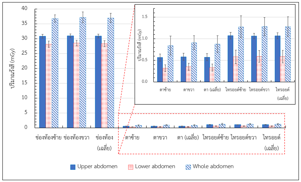
Background: Abdominal computed tomography (CT) plays a crucial role in a diagnosis of disease and continually increases. However, X-ray radiation is harmful and requires a careful monitoring of patient radiation dose obtained from this procedure. Objectives: This study aimed to determine patient radiation dose from upper abdomen CT, lower abdomen CT and whole abdomen CT at Maharat Nakhon Ratchasima Hospital and compare this data with national diagnostic reference levels (NDRLs). Moreover, the evaluation of entrance surface air kerma (ESAK) of abdomen, eyes, and thyroid were performed. Methods: Patients who had undergone upper abdomen CT, lower abdomen CT, and whole abdomen CT (30 patients for each examination) between February and April 2023, and met the inclusion criteria, were recruited. The nanoDot optically stimulated luminescence dosimeters (OSLDs) were placed on patient’s skin at the eyes, thyroid, and abdomen to measure the ESAK. The radiation dose parameters including volumetric CT dose index (CTDIvol), dose length product (DLP), total DLP were recorded. Furthermore, an effective dose was calculated. Results: The highest values of CTDIvol (13.45 mGy), DLP (714.19 mGy.cm), total DLP (2575.20 mGy.cm), and effective dose (38.50 mSv) were obtained from the whole abdomen CT examination. However, CTDIvol and DLP values for upper abdomen CT and whole abdomen CT were lower than the NDRLs. The highest ESAK values were consequently observed for the eyes (0.88 ± 0.19 mGy), thyroid (1.28 ± 0.23 mGy), and abdomen (36.98 ± 1.54 mGy) for the whole abdomen CT examination. Conclusion: The CTDIvol and DLP values obtained from abdominal CT at Maharat Nakhon Ratchasima Hospital were within the NDRLs, indicating that our abdominal CT protocols for both upper abdomen and whole abdomen can be used to examine patients appropriately.
Downloads
References
OCED. Computed tomography (CT) exams (indicator). doi:10.1787/3c994537-en. Accessed on 1 August 2023. https://data.oecd.org/healthcare/computed-tomography-ct-exams.htm
Poosiri S, Krisanachinda A, Khamwan K. Evaluation of patient radiation dose and risk of cancer from CT examinations. Radiol Phy Technol. 2023. https://dol.org/10.1007/s12194-023-00763-w.
Berrington de GA, Darby S. Risk of cancer from diagnostic X-rays: estimates for the UK and 14 other countries. Lancet. 2004;363(9406):345-51.
Vano E, Miller DL, Martin CJ, Rehani MM, Kang K, Rosenstein M, et al. ICRP Publication 135: Diagnostic Reference Levels in Medical Imaging. Ann ICRP. 2017;46(1):1-44. doi: 10.1177/0146645317717209.
Kanal KM, Butler PF, Sengupta D, Bhargavan-Chatfield M, Coombs LP, Morin RL. U.S. Diagnostic Reference Levels and Achievable Doses for 10 Adult CT Examinations. Radiology. 2017;284(1):120-33.
กรมวิทยาศาสตร์การแพทย์ กระทรวงสาธารณสุข. ค่าปริมาณรังสีอ้างอิงในการถ่ายภาพรังสีวินิจฉัยทางการแพทย์ของประเทศไทย 2564. กรุงเทพมหานคร: บริษัทบียอนด์พับลิสซิ่ง จำกัด; 2564.
Pema D, Kritsaneepaiboon S. Radiation Dose from Computed Tomography Scanning in Patients at Songklanagarind Hospital: Diagnostic Reference Levels. J Health Sci Med Res. 2020;38(2):135-43.
วลัยรัตน์ เงาดีงาม. ปริมาณรังสีที่ผู้ป่วยได้รับจากการตรวจด้วยเอกซเรย์คอมพิวเตอร์ของโรงพยาบาลสิชล. วารสารโรงพยาบาลนครพิงค์. 2564;12(2):97-107.
Donmoon T, Chusin T. Establishment of local diagnostic reference levels for commonly performed computed tomography examinations in Thai cancer hospitals. Thai J Rad Tech. 2021;46(1):35-42.
Sodkokruad P, Asvaphatiboon S, Thanabodeebonsiri J, Tangboonduangit P. Comparision of computed tomography dose index measuring by two detector types of computed tomography simulator. Thai J Rad Tech. 2018;43(1):64-8.
Virginia TS, John EA, Raju S, Maria AS, Anchali K, Madan R, et al. Dose reduction in CT while maintaining diagnostic confidence: diagnostic reference levels at routine head, chest and abdominal CT-IAEA-coordinated research project. Radiology. 2006;240:828-34.
เขมิกา เกื้อพิทักษ์, จุฑามาศ ทนานนท์, ปนัสดา อวิคุณประเสริฐ, สุนิสา แสงสว่าง, จิรภัทร เรืองศรีตระกูล, วิทิต ผึ่งกัน, และคณะ. การศึกษาปริมาณรังสีและปริมาณรังสีกระเจิงจากการตรวจด้วยเครื่องเอกซเรย์คอมพิวเตอร์ในหุ่นจำลอง. J Med Health Sci. 2019;26:19-28.
Nupetch S, Awikunprasert P, Pungkun V. Radiation dose response of Inlight® optically stimulated luminescence (OSL) dosimeter. Thai J Rad Tech. 2018;43(1):36-43.
กรมวิทยาศาสตร์การแพทย์ กระทรวงสาธารณสุข. ค่าปริมาณรังสีอ้างอิงในการถ่ายภาพรังสีวินิจฉัยทางการแพทย์ของประเทศไทย 2566. กรุงเทพมหานคร: บริษัทบียอนด์พับลิสซิ่ง จำกัด; 2566.
Pimsorn P. Factor affecting size–specific dose estimates in addition to scanning parameters for chest–abdomen–pelvis computed tomography using automatic tube current modulation at Chiangrai prachanukroh hospital. Thai J Rad Tech. 2022 Dec.31; (1):43-54.
Allisy-Roberts P, Williams J. Radiation physics. W.B. Saunders; 2008. p. 1–21. Available from: https://www.sciencedirect.com/science/article/pii/B9780702028441500053.
Antonio PL, Caldas LVE. Angular dependence of Tl and OSL responses of Al2O3:C commercial detectors in standard beta radiation beams. Radiat. Prot. Dosim. 2014, 113, 359–365.
Okazaki T, Hayashi H, Takegami K, Okino H, Kimoto N, Maehata I, et al. Fundamental Study of NanoDot OSL Dosimeters for Entrance Skin Dose Measurement in Diagnostic X-Ray Examinations. J. Radiat Prot. Res. 2016, 41, 229–236.
Jursinic PA. Optically stimulated luminescent dosimeters stable response to dose after repeated bleaching. Med Phys. 2020 Jul;47(7):3191-3203.
Zhuang AH, Olch AJ. A practical method for the reuse of nanoDot OSLDs and predicting sensitivities up to at least 7000 cGy. Med Phys. 2020 Apr;47(4):1481-1488.
Kanal KM, Butler PF, Sengupta D, Bhargavan-Chatfield M, Coombs LP, Morin RL. U.S. diagnostic reference levels and achievable doses for 10 adult CT examinations. Radiology. 2017;284(1):120-33.
Radiation Protection No 180. Diagnostic reference levels in thirty-six European countries [monograph on the Internet]. European Commission (EC). Luxembourg: Publication Office of the European Union; 2014 [cited 2019 Feb 11]. Available from: http://ec.europa>files>RP180 part2.
National Diagnostic Reference Levels in Japan (2020) Japan: Japan Network for Research and Information on Medical Exposure (J-RIME); 2020. Available from: https://www.jsmp.org/en/info/national-diagnostic-reference-levels-in-japan-2020-has-been-released/.
Hayton A, Wallace A, Marks P, Edmonds K, Tingey D, Johnston P. Australian diagnostic reference levels for multi-detector computed tomography. Australas Phys Eng Sci Med. 2013;36:19-26.
Trinavarat P, Kritsaneepaiboon S, Rongviriyapanich C, Visrutaratna P, Srinakarin J. Radiation dose from CT scanning: can it be reduced? Asian Biomedicine. 2011;5:13-21.
Nuntue C, Krisanachinda A, Khamwan K. Optimization of a low-dose 320-slice multidetector computed tomography chest protocol using a phantom. Asian Biomedicine. 2016;10:269-276.
Downloads
Published
How to Cite
Issue
Section
License
Copyright (c) 2023 The Thai Society of Radiological Technologists

This work is licensed under a Creative Commons Attribution-NonCommercial-NoDerivatives 4.0 International License.
บทความที่ได้รับการตีพิมพ์เป็นลิขสิทธิ์ของสมาคมรังสีเทคนิคแห่งประเทศไทย (The Thai Society of Radiological Technologists)
ข้อความที่ปรากฏในบทความแต่ละเรื่องในวารสารวิชาการเล่มนี้เป็นความคิดเห็นส่วนตัวของผู้เขียนแต่ละท่านไม่เกี่ยวข้องกับสมาคมรังสีเทคนิคแห่งประเทศไทยและบุคคลากรท่านอื่น ๆในสมาคม ฯ แต่อย่างใด ความรับผิดชอบองค์ประกอบทั้งหมดของบทความแต่ละเรื่องเป็นของผู้เขียนแต่ละท่าน หากมีความผิดพลาดใดๆ ผู้เขียนแต่ละท่านจะรับผิดชอบบทความของตนเองแต่ผู้เดียว


