
The outcome of exposure technique adjustment toward the deviation index and national diagnostic reference levels from digital chest radiographs in Lomkao Crown Prince Hospital
Keywords:
Exposure technique for digital chest radiographs, Deviation index, Radiographic chest x-rayAbstract
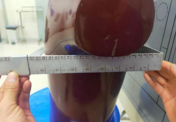
Introduction: The Deviation Index (DI) plays a crucial role in digital imaging systems, guiding the selection of optimal radiographic techniques. Objective: This study aimed to optimize the chest postero-anterior (PA) X-ray radiographic technique with the optimal DI value as per AAPM recommendations, ensuring that the Entrance Skin Air Kerma (ESAK) stays within specified exposure limits. Furthermore, the study sought to compare the DI values before and after adjusting the exposure techniques. Methods: The radiographic parameters for chest PA X-ray were optimized by varying kVp and mAs while exposing a phantom. The ESAK and DI were recorded for each combination of exposure techniques. An appropriate exposure technique, according to the AAPM standard that determines the DI criteria which to be between ±3 and an ESAK value not exceeding 0.4 mGy, was selected after the adjustment was performed. The original and adjusted exposure techniques were then applied to two patient groups, each consisting of 34 patients. Subsequently, a comparison was made between the original and the adjusted technique values. Results: The default exposure technique was 117.41±1.51 kVp and 3.7±0.76 mAs, resulting in chest radiographs with an average DI of -4.11±0.91 and an ESAK value at the median of 0.22 mGy. After adjusting the exposure technique to 124 kVp and 4.5 mAs, the average DI was -2.38±0.53, and the ESAK value at the median was 0.27 mGy. When comparing the average DI values and the percentage of images meeting AAPM standards before and after modifying the exposure technique, a statistically significant difference was observed (p-value<0.001). The number of radiographs with DI values within the AAPM standard increased to 85% of the total images, compared to 8% with the default exposure technique. Conclusion: The adjusted radiographic technique for chest radiographs achieves DI values within the standard criteria of AAPM and keeps ESAK values below the suggested reference level (Thailand DRLs 2023). Furthermore, the image quality is superior to that of the original technique. Therefore, this study recommends the adoption of the adjusted radiographic technique for digital chest radiography.
Downloads
References
Butler ML, Rainford L, Last J, Brennan PC. Are exposure index values consistent in clinical practice? A multi-manufacturer investigation. Radiat Prot Dosimetry. 2010;139(1–3):371–374.
Lança L, Silva A. Evaluation of exposure index (IgM) in orthopaedic radiography. Radiat Prot Dosimetry. 2008;129(1–3):112–128.
Shepard SJ, Wang J, Flynn M, Gingold E, Goldman L, Krugh K, et al. An exposure indicator for digital radiography: AAPM Task Group 116 (executive summary). Med Phys. 2009;36(7):2898–2914.
Don S, Whiting BR, Rutz LJ, Apgar BK. New exposure indicators for digital radiography simplified for radiologists and technologists. AJR Am J Roentgenol. 2012;199(6):1337–1341.
ประภัสสร ไกรหาญ, ดวงฤดี สุภมาตย์. การประเมินค่าดัชนีชี้วัดปริมาณรังสีในการถ่ายภาพทรวงอกเด็กอายุ 0-12 ปี โรงพยาบาลสิรินธร จังหวัดขอนแก่น. วารสารสุขภาพและสิ่งแวดล้อมศึกษา. 2564;4:18–27.
กรมวิทยาศาสตร์การแพทย์ กระทรวงสาธารณสุข. ค่าปริมาณรังสีอ้างอิงจากการถ่ายภาพรังสีวินิจฉัยจากเครื่องเอกซเรย์ทั่วไปแบบดิจิตอล 2566 (Diagnostic reference levels in Digital radiography: DR). กรุงเทพฯ: บียอนด์ พับสิสชิ่ง; 2566.
วิทยาศาสตร์และเทคโนโลยีการกีฬา มหาวิทยาลัยมหิดล. คนไทยรู้ยัง: คนไทยวัยทำงานมี “ดัชนีมวลกายปกติ” แค่ 51.76% [อินเทอร์เน็ต]. 2562 [เข้าถึงเมื่อ 20 ก.พ. 2566]. เข้าถึงได้จาก: https://www.tcijthai.com/news/2019/13/scoop/9662
Daniel WW, Cross CL. Foundations for analysis in the health sciences. Biom J. 1995;37(6):177–178.
สุรพงษ์ คงสัตย์, ธีรชาติ ธรรมวงค์. การหาค่าความเที่ยงตรงของแบบสอบถาม (IOC) [อินเทอร์เน็ต]. 2551 [เข้าถึงเมื่อ 22 มี.ค. 2566]. เข้าถึงได้จาก: https://www.mcu.ac.th/article/detail/14329
Vañó E, Miller DL, Martin CJ, Rehani MM, Kang K, Rosenstein M, et al. ICRP publication 135: Diagnostic reference levels in medical imaging. Ann ICRP. 2017;46(1):111–144.
International Atomic Energy Agency. Dosimetry in diagnostic radiology: an international code of practice. IAEA Technical Reports Series No. 457. Vienna: IAEA; 2007.
วสุชาวลี เชื้อมหาวัน, ศิริวรรณ บุญชรัตน. คู่มือการวัดค่าปริมาณรังสีที่ใช้ในการถ่ายภาพรังสีด้วยเครื่องเอกซเรย์ [อินเทอร์เน็ต]. 2566 [เข้าถึงเมื่อ 30 กันยายน 2566]. เข้าถึงได้จาก: https://webapp1.dmsc.moph.go.th/petitionxray/web3/downloadproject/รายละเอียดโครงการและวิธีการเก็บข้อมูล.pdf
เสาวนีย์ อัศวผาติบุญ. การป้องกันอันตรายจากรังสีวินิจฉัย [อินเทอร์เน็ต]. 2561 [เข้าถึงเมื่อ 30 ก.ย. 2566]. เข้าถึงได้จาก: https://www.rama.mahidol.ac.th/radiology/sites/default/files/public/knowledge/A_Diagnosis_Protection_24July2018_handout.pdf
Japan Association on Radiological Protection in Medicine. Diagnostic reference levels based on latest surveys in Japan [Internet]. 2015 [cited 29 Sept 2023]. Available from: http://www.radher.jp/J-RIME/report/DRLhoukokusyoEng.pdf
คณะแพทยศาสตร์ มหาวิทยาลัยมหิดล. Radiation protection in diagnostic radiology 2018 [อินเทอร์เน็ต]. 2561 [เข้าถึงเมื่อ 30 กันยายา 2566]. เข้าถึงได้จาก:
https://www.rama.mahidol.ac.th/radiology/sites/default/files/public/training/Protection2018.pdf
รังสีวิทยาสมาคมแห่งประเทศไทย. โอกาสเกิดมะเร็งกับการตรวจวินิจฉัยทางรังสี [อินเทอร์เน็ต]. 2562 [เข้าถึงเมื่อ 16 ส.ค. 2566]. เข้าถึงได้จาก: https://www.radiologythailand.org/
ลัดดา เย็นศรี. การศึกษาเปรียบเทียบปริมาณรังสีที่ผู้ป่วยได้รับจากการถ่ายภาพรังสีทรวงอกด้วยระบบ CR และ DR. วารสารเครือข่ายวิทยาลัยพยาบาลและการสาธารณสุขภาคใต้. 2559;3(1):129–139.
ธัญรัตน์ ชูศิลป์, ฐิติพงศ์ แก้วเหล็ก. ตัวบ่งชี้ปริมาณรังสีในระบบการถ่ายภาพรังสีแบบดิจิทัล. วารสารรังสีเทคนิค. 2561;43(1):21–28.
Downloads
Published
How to Cite
Issue
Section
License
Copyright (c) 2024 The Thai Society of Radiological Technologists

This work is licensed under a Creative Commons Attribution-NonCommercial-NoDerivatives 4.0 International License.
บทความที่ได้รับการตีพิมพ์เป็นลิขสิทธิ์ของสมาคมรังสีเทคนิคแห่งประเทศไทย (The Thai Society of Radiological Technologists)
ข้อความที่ปรากฏในบทความแต่ละเรื่องในวารสารวิชาการเล่มนี้เป็นความคิดเห็นส่วนตัวของผู้เขียนแต่ละท่านไม่เกี่ยวข้องกับสมาคมรังสีเทคนิคแห่งประเทศไทยและบุคคลากรท่านอื่น ๆในสมาคม ฯ แต่อย่างใด ความรับผิดชอบองค์ประกอบทั้งหมดของบทความแต่ละเรื่องเป็นของผู้เขียนแต่ละท่าน หากมีความผิดพลาดใดๆ ผู้เขียนแต่ละท่านจะรับผิดชอบบทความของตนเองแต่ผู้เดียว


