Optimizing the minimum detectable difference of chest protocol digital radiography system by V-shaped line gauge phantom and Taguchi analysis
Keywords:
V-shaped line gauge phantom, Minimum Detectable Difference, Taguchi analysis, Digital RadiographyAbstract
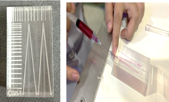
Background: The Minimum Detectable Difference (MDD) of Digital Radiography (DR) image systems was quantified and optimized using a self-developed V-shaped line gauge phantom and Taguchi optimization analysis. The increased clinical applications of the DR system are mainly due to its ability to provide instant and precise imaging for radiologists to diagnose. Objective: The purpose of this study was to optimize parameters for MDD values based on Taguchi's analysis and to verify a self-developed V-shaped line gauge phantom for the chest protocol of the DR system. Materials and methods: The chest protocol was exposed using the V-shaped line gauge and solid acrylic water phantom in various sizes with the DR system. Five factors were assigned in this study: (A) focal spot, (B) kVp, (C) mAs, (D) filter, and (E) phantom thickness. Since each factor could have two or three levels, eighteen groups of factor combinations were organized according to Taguchi’s algorithm. The V-shaped line gauge images were estimated. ANOVA was adopted to determine the significant factors and MDD values to evaluate the spatial resolution. Results: The optimal parameters for the highest Signal-to-Noise (S/N) ratio were as follows: (A) large focal spot, (B) 140 kVp, (C) 6.25 mAs, (D) 0.1 mmCu filter, and (E) phantom thickness of 16 cm. Conclusion: The optimal parameters in this study provided an MDD value of 0.87 mm compared to 1.66 mm for conventional parameters. The self-developed V-shaped line gauge phantom also proved to be suitable for the DR system. Furthermore, all factors affected the image quality with statistical significance following the ANOVA analysis.
Downloads
References
Krishnaiah K, Shahabudeen P. Fundamentals of experimental design. In: Ghosh AK, editors. Applied design of experiments and Taguchi methods. New Delhi: PHI Learning; 2012;22-48.
Krishnaiah K, Shahabudeen P. Taguchi methods. In: Ghosh AK, editors. Applied design of experiments and Taguchi methods. New Delhi: PHI Learning; 2012;198-201.
พรเพ็ญ รุจิรามงคลชัย, ภัทรภณ หนูแก้ว. การหาค่าพารามิเตอร์ที่เหมาะสมในการถ่ายภาพด้วยเครื่องเอกซเรย์ DR สำหรับเทคนิคการถ่ายภาพทรวงอกโดยอาศัยการวิเคราะห์ของทากูชิ: ศึกษาจากหุ่นจำลอง [ภาคนิพนธ์ปริญญาวิทยาศาสตรบัณฑิต]. นครปฐม: มหาวิทยาลัยมหิดล; 2561.
วสวัตติ์ ประสงค์สร้าง, สิรัณยาพงศ์ สุวรรณโอภาส. ข้อแตกต่างระหว่างการเอกซเรย์ระบบถ่ายภาพรังสีคอมพิวเตอร์กับระบบถ่ายภาพดิจิทัล. วารสารรังสีวิทยาศิริราช 2018;5(1):48-54.
โรงพยาบาลสำโรงการแพทย์. เครื่องเอกซเรย์ระบบดิจิตอล [อินเทอร์เน็ต]. 2555 [เข้าถึงเมื่อ 27 ธ.ค. 2565]. เข้าถึงได้จาก: http://www.samrong-hosp.com/ข่าวและกิจกรรม/เครื่องเอกซเรย์ระบบดิจิตอล
Pan LF, Wu KY, Chen KL, Kittipayak S, Pan LK. Taguchi method-based optimization of the minimum detectable difference of a cardiac x-ray imaging system using a precise line pair gauge. J Mech Med Biol 2019;19(7):1-17.
Yeh DM, Chang PJ, Pan LK. The optimum Ga-67-citrate gamma camera imaging quality factors as first calculated and shown by the Taguchi’s analysis. Hell J Nucl Med 2013;16(1):25-32.
Pan LF, Chen YH, Wang CC, Peng BR, Kittipayak S, Pan LK. Optimizing cardiac CT angiography minimum detectable difference via Taguchi’s dynamic algorithm, a V-shaped line gauge, and three PMMA phantoms. Technol Health Care 2022; 30:91–103.
สุชาติ เกียรติวัฒนเจริญ. kV หรือ kVp [อินเทอร์เน็ต]. 2553 [เข้าถึงเมื่อ 2 ก.พ. 2566]. เข้าถึงได้จาก: https://www.gotoknow.org/posts/401227
Bell DJ. Milliampere-seconds (mAs) [Internet]. 2021 [cited 2023 February 2]. Available from: https://radiopaedia.org/articles/milliampere-seconds-mas45
เพชรากร หาญพานิชย์. การกรองรังสีของหลอดเอกซเรย์ [อินเทอร์เน็ต]. 2553 [เข้าถึงเมื่อ 2 ก.พ. 2566]. เข้าถึงได้จาก: https://www.gotoknow.org/posts/
Supertech x-ray. Whole Body Phantom Kyoto Kagaku PBU-60 [Internet]. 2022 [cited 2022 October 19]. Available from: https://www.supertechx-ray.com/Anthropomorp hic/FullBodyPhantoms/PBU-60.php#
Larson MG. Analysis of Variance. Circulation 2008;117(1):115-121.
Chieng R, Alhusseiny K, Liao A. Spatial resolution (gamma camera) [Internet]. 2023 [cited 2024 April 3]. Available from: https://doi.org/10.53347/rID-173540

Downloads
Published
How to Cite
Issue
Section
License
Copyright (c) 2024 The Thai Society of Radiological Technologists

This work is licensed under a Creative Commons Attribution-NonCommercial-NoDerivatives 4.0 International License.
บทความที่ได้รับการตีพิมพ์เป็นลิขสิทธิ์ของสมาคมรังสีเทคนิคแห่งประเทศไทย (The Thai Society of Radiological Technologists)
ข้อความที่ปรากฏในบทความแต่ละเรื่องในวารสารวิชาการเล่มนี้เป็นความคิดเห็นส่วนตัวของผู้เขียนแต่ละท่านไม่เกี่ยวข้องกับสมาคมรังสีเทคนิคแห่งประเทศไทยและบุคคลากรท่านอื่น ๆในสมาคม ฯ แต่อย่างใด ความรับผิดชอบองค์ประกอบทั้งหมดของบทความแต่ละเรื่องเป็นของผู้เขียนแต่ละท่าน หากมีความผิดพลาดใดๆ ผู้เขียนแต่ละท่านจะรับผิดชอบบทความของตนเองแต่ผู้เดียว


