
Comparison of F-18 FDG standardized uptake value in PET images attenuation-corrected using CT data with and without contrast media in head and neck cancer
Keywords:
SUV, PET images, CT data, Contrast media, Attenuation correctionAbstract
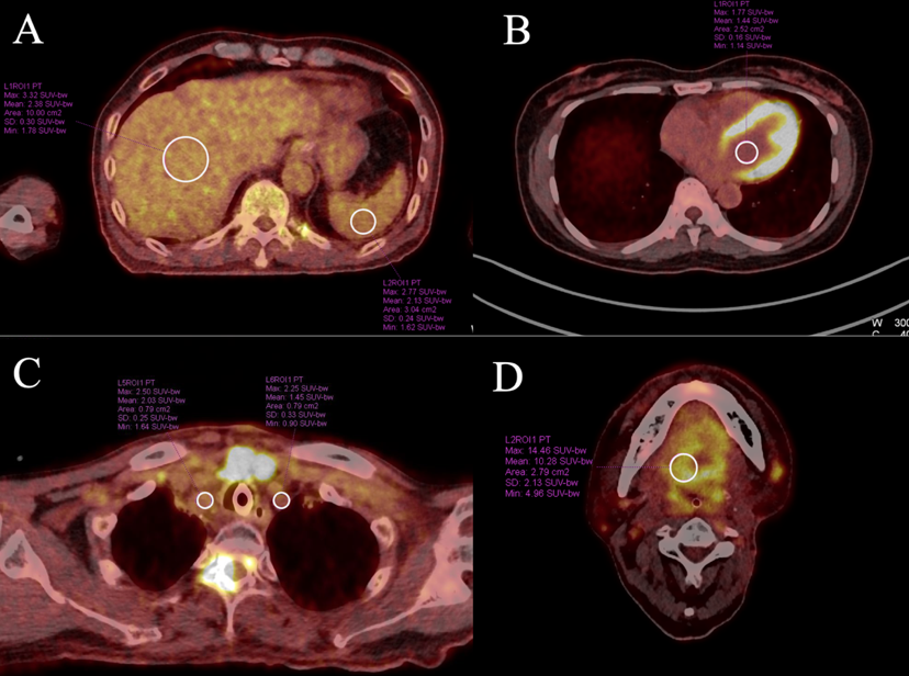
Background: PET and CT scanners are now integrated into a single system, enabling the combination of PET images, which display the functional activity of organs, with CT images, which provide detailed anatomical information. Moreover, CT images can be used for attenuation correction of PET data, as they reflect the tissue's radiation absorption properties in regions where photons are present. However, when CT data acquired with contrast media is used for PET image reconstruction, attenuation correction values may be overestimated. This can result in inaccuracies in the evaluation of the Standardized Uptake Value (SUV). Objective: To compare the SUV from PET images corrected for attenuation using CT data with and without contrast media in head and neck cancer patients. Methods: The study was conducted using a phantom containing F-18 FDG at 157 MBq, with three different volumes of contrast media: 0, 100, and 200 milliliters. Regions of interest (ROIs) were drawn in five locations with uniform radioactivity distribution. Subsequently, a clinical study was performed on 32 patients, with a mean age of 48.33 ± 13.27 years, all of whom had no liver abnormalities. ROIs were drawn in five areas: the liver, spleen, heart, subclavian vein, and tumor mass. Results: The percentage differences in SUV values obtained from PET images with attenuation correction using CT data with contrast media, compared to those without contrast media, in the liver, spleen, heart, subclavian vein, and tumor mass were as follows: SUVmax of 7.64 ± 4.19%, 8.05 ± 5.17%, 12.32 ± 5.73%, 9.55 ± 4.85%, and 7.09 ± 5.51%, respectively. The percentage differences in SUVmean values were 8.19 ± 5.34%, 8.68 ± 5.70%, 12.54 ± 5.86%, 10.04 ± 4.97%, and 6.01 ± 5.04%, respectively. These differences were statistically significant (P value < 0.05) and showed a strong linear correlation between the two correction methods (R² > 0.95). Conclusion: The SUVmax and SUVmean values from PET images corrected for attenuation using CT data with contrast agent differed significantly from those without contrast.
Downloads
References
Townsend DW, Carney JPJ, Yap JT, Hall NC. PET/CT Today and Tomorrow. J Nucl Med. 2004;45(1):4S-14S.
Bar-Shalom R, Yefremov N, Guralnik L, Gaitini D, Frenkel A, Kuten A, et al. Clinical performance of PET/CT in evaluation of cancer: additional value for diagnostic imaging and patient management. J Nucl Med. 2003;44(8):1200-9.
Koshy M, Paulino AC, Howell R, Schuster D, Halkar R, Davis LW. F-18 FDG PET-CT fusion in radiotherapy treatment planning for head and neck cancer. Head Neck. 2005;27(6):494-502.
Crișan G, Moldovean-Cioroianu NS, Timaru DG, Andrieș G, Căinap C, Chiș V. Radiopharmaceuticals for PET and SPECT Imaging: A Literature Review over the Last Decade. Int J Mol Sci. 2022;23(9):5023.
Trotter J, Pantel AR, Teo BKK, Escorcia FE, Li T, Pryma DA, et al. Positron Emission Tomography (PET)/Computed Tomography (CT) Imaging in Radiation Therapy Treatment Planning: A Review of PET Imaging Tracers and Methods to Incorporate PET/CT. Adv Radiat Oncol. 2023 8(5):101212.
Khamwan K, Krisanachinda A, Pasawang P. The determination of patient dose from 18F-FDG PET/CT examination. Radiat Prot Dosimetry. 2010 Sep;141(1):50-5.
Khamwan K, O'Reilly SE, Plyku D, Goodkind A, Josefsson A, Cao X, Fahey FH, Treves ST, Bolch WE, Sgouros G. Re-evaluation of pediatric 18F-FDG dosimetry: Cristy-Eckerman versus UF/NCI hybrid computational phantoms. Phys Med Biol. 2018 Aug 14;63(16):165012.
Wongsa P, Panperee P, Wangrattanaampai J, Piantham W, Wongkai C, Jantarato A. A comparison of efficiency between 18F-FDG PET neuroimaging analysis software in Alzheimer’s disease patients. Thai J Rad Tech;47(1):64-72.
Israel O, Mor M, Gaitini D, Keidar Z, Guralnik L, Engel A, et al. Combined functional and structural evaluation of cancer patients with a hybrid camera-based PET/CT system using 18F-FDG. J Nucl Med. 2002;43(9):1129-36.
Treglia G, Sadeghi R, Sole AD, Giovanella L. Diagnostic performance of PET/CT with tracers other than F-18-FDG in oncology: an evidence-based review. Clin Transl Oncol. 2014 16(9):770-5.
Boellaard R, Delgado-Bolton R, Oyen WJG, Giammarile F, Tatsch K, Eschner W, et al. FDG PET/CT: EANM procedure guidelines for tumour imaging: version 2.0. Eur J Nucl Med Mol Imaging. 2015;42(2):328-54.
Yau YY, Chan WS, Tam YM, Vernon P, Wong S, Coel M, et al. Application of intravenous contrast in PET/CT: does it really introduce significant attenuation correction error? J Nucl Med. 2005;46(2):283-91.
Kinahan PE, Fletcher JW. PET/CT Standardized Uptake Values (SUVs) in Clinical Practice and Assessing Response to Therapy. Seminars in ultrasound, CT, and MR. 2010;31(6):496–505.
Mawlawi O, Erasmus JJ, Munden RF, Pan T, Knight AE, Macapinlac HA, et al. Quantifying the effect of IV contrast media on integrated PET/CT: clinical evaluation. AJR Am J Roentgenol. 2006;186(2):308-19.
ter Voert EE, van Laarhoven HW, Kok PJ, Oyen WJ, Visser EP, de Geus-Oei LF. Comparison of liver SUV using unenhanced CT versus contrast-enhanced CT for attenuation correction in 18F-FDG PET/CT. Nucl Med Commun. 2014 May;35(5):472-7.
Rebière M, Verburg FA, Palmowski M, Krohn T, Pietsch H, Kuhl CK, et al. Multiphase CT scanning and different intravenous contrast media concentrations in combined F-18-FDG PET/CT: Effect on quantitative and clinical assessment. Eur J Radiol. 2012;81(8):e862-e9.
Downloads
Published
How to Cite
Issue
Section
License
Copyright (c) 2025 The Thai Society of Radiological Technologists

This work is licensed under a Creative Commons Attribution-NonCommercial-NoDerivatives 4.0 International License.
บทความที่ได้รับการตีพิมพ์เป็นลิขสิทธิ์ของสมาคมรังสีเทคนิคแห่งประเทศไทย (The Thai Society of Radiological Technologists)
ข้อความที่ปรากฏในบทความแต่ละเรื่องในวารสารวิชาการเล่มนี้เป็นความคิดเห็นส่วนตัวของผู้เขียนแต่ละท่านไม่เกี่ยวข้องกับสมาคมรังสีเทคนิคแห่งประเทศไทยและบุคคลากรท่านอื่น ๆในสมาคม ฯ แต่อย่างใด ความรับผิดชอบองค์ประกอบทั้งหมดของบทความแต่ละเรื่องเป็นของผู้เขียนแต่ละท่าน หากมีความผิดพลาดใดๆ ผู้เขียนแต่ละท่านจะรับผิดชอบบทความของตนเองแต่ผู้เดียว


