
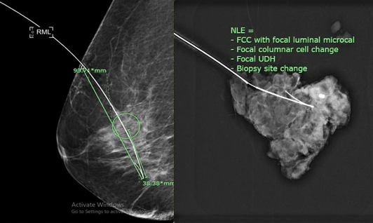
Needle localization under mammographic guidance
Keywords:
Mammography, Breast cancer, Needle localization, Breast cancer screeningAbstract
Within the medical history of cancer, breast cancer is a prevalent form of cancer affecting Thai women and globally. To enhance the survival rates of breast cancer patients, it is crucial to undergo regular annual mammogram screenings. Early detection of lesions facilitates prompt initiation of treatment. Surgical intervention remains the most effective treatment method for patients whose lesions are detected at an early stage. This is particularly true for non-palpable lesions or those with ill-defined margins. In such cases, needle localization techniques become necessary to precisely determine the lesion’s location prior to surgical intervention. This facilitates accurate surgical procedure and comprehensive lesion removal. The choice of needle localization technique depends on the lesion’s location and the availability of specific instrument at the healthcare facility. This article provides a comprehensive overview and description of needle localization for lesion identification under mammographic guidance. It encompasses the indications for the procedure, including two-dimension and three-dimension mammographic techniques, the requisite devices and needle techniques employed, potential limitations, and associated patient risks during the procedure.
Downloads
References
American Cancer Society. Breast cancer statistics | How common is breast cancer? [Internet]. 2023 [cited 2024 Aug 27]. Available from: https://www.cancer.org/cancer/types/ breast-cancer/about/how-common-is-breast-cancer.html
Kalambo M, Dogan BE, Whitman GJ. Step by step: planning a needle localization procedure. Clin Imaging. 2020;60:100-8.
Verkooijen HM, Peeters PHM, Pijnappel RM, Koot VCM, Schipper MEI, Rinkes IHMB. Diagnostic accuracy of needle-localized open breast biopsy for impalpable breast disease. Br J Surg. 2000;87:344-7.
Namarak Hospital. ผ่าตัดก้อนเต้านม [Internet]. 2024 [cited 2024 Aug 27]. Available from: https://www.namarak.com/breast/detail/3/17
Guirguis MS, Adrada BE, Scoggins ME, Moseley TW, Dryden MJ, Le-Petross HC, et al. The challenging image-guided preoperative breast localization: a modality-based approach. AJR Am J Roentgenol. 2022;218:423-34.
Shin K, Teichgraeber D, Martaindale S, Whitman GJ. Tomosynthesis-guided core biopsy of the breast: why and how to use it. J Clin Imaging Sci. 2018;8:1-5.
Choudhery S, Simmons C, Harper L, Lee CU. Tomosynthesis-guided needle localization of breast and axillary lesions: our initial experience. AJR Am J Roentgenol. 2019;212:943-6.
Songsaeng C, Modmontin K, Punubon K. Local diagnostic reference levels for breast screening using digital mammography at Tanyawej Breast Center, Songklanagarind Hospital. Thai J Rad Tech. 2020;44(1):1-7.
Chatchoedtragoon C, Sriprasert N, Tuasakul S, Kaewlek T. Development of breast lesion detection program. Thai J Rad Tech. 2021;45(1):8-12.
Davies H. Digital tomosynthesis (3D mammography) [Internet]. EBME. [cited 2024 Aug 27]. Available from: https://www.ebme.co.uk/articles/clinical-engineering/3d-mammography
Fujifilm. AMULET Innovality: advanced biopsy system [Internet]. 2024 [cited 2024 Aug 24]. Available from: https://www.fujifilm.com/sg/en/healthcare/x-ray/mammography/amulet-innovality/advanced-biopsy-system
Liberman L. Percutaneous image-guided core breast biopsy. Radiol Clin North Am. 2002;40(3):483-500.
American College of Radiology. ACR practice parameter for the performance of stereotactic-guided breast interventional procedures. Reston (VA): American College of Radiology; 2016.
Downloads
Published
How to Cite
Issue
Section
License
Copyright (c) 2025 The Thai Society of Radiological Technologists

This work is licensed under a Creative Commons Attribution-NonCommercial-NoDerivatives 4.0 International License.
บทความที่ได้รับการตีพิมพ์เป็นลิขสิทธิ์ของสมาคมรังสีเทคนิคแห่งประเทศไทย (The Thai Society of Radiological Technologists)
ข้อความที่ปรากฏในบทความแต่ละเรื่องในวารสารวิชาการเล่มนี้เป็นความคิดเห็นส่วนตัวของผู้เขียนแต่ละท่านไม่เกี่ยวข้องกับสมาคมรังสีเทคนิคแห่งประเทศไทยและบุคคลากรท่านอื่น ๆในสมาคม ฯ แต่อย่างใด ความรับผิดชอบองค์ประกอบทั้งหมดของบทความแต่ละเรื่องเป็นของผู้เขียนแต่ละท่าน หากมีความผิดพลาดใดๆ ผู้เขียนแต่ละท่านจะรับผิดชอบบทความของตนเองแต่ผู้เดียว


