
การวิเคราะห์การถ่ายฟิล์มซ้ำและผลกระทบต่อการประกันคุณภาพในงานรังสีวินิจฉัย: กรณีศึกษาในสถาบัน
คำสำคัญ:
การวิเคราะห์อัตราการถ่ายเอกซเรย์ซ้ำ, การวิเคราะห์การปฏิเสธและการใช้งาน, ภาพถ่ายเอกซเรย์ทรวงอก, การประกันคุณภาพในงานรังสีวินิจฉัยบทคัดย่อ
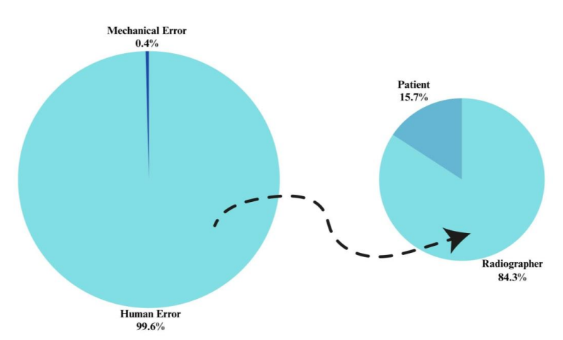
งานวิจัยนี้มีวัตถุประสงค์เพื่อวิเคราะห์อัตราการตรวจทรวงอกซ้ำโดยใช้โปรแกรมเครื่องมือวิเคราะห์การปฏิเสธและการใช้งาน (Reject and Usage Analysis) ของเครื่องเอกซเรย์ Samsung XGEO-GC80 การศึกษาวิจัยเป็นแบบย้อนหลัง (Retrospective study) ดำเนินการที่แผนกรังสีวินิจฉัย โรงพยาบาลจุฬาลงกรณ์ สภากาชาดไทย การศึกษานี้เกี่ยวข้องกับการรวบรวมสถิติและตรวจสอบสาเหตุที่อยู่เบื้องหลังรูปภาพที่ถูกปฏิเสธภายในระบบเครื่องเอกซเรย์อย่างละเอียด ผลการวิจัยที่สำคัญ ได้แก่ การถ่ายภาพเอกซเรย์ทรวงอกมีจำนวนภาพมากที่สุด คิดเป็น 39.32% ของภาพทั้งหมด 40,400 ภาพ ภาพที่มีการถ่ายซ้ำที่พบได้บ่อยที่สุดในการถ่ายภาพเอกซเรย์ทรวงอกคิดเป็น 32.21% จาก 7,550 ภาพที่ถูกปฏิเสธ และ 6.02% ของภาพทั้งหมด สาเหตุหลักในการปฏิเสธภาพ คือการวางตำแหน่งจัดท่าที่ไม่เหมาะสม คิดเป็น 75.47% ของภาพที่ถูกปฏิเสธทั้งหมด สาเหตุที่พบบ่อยที่สุดสำหรับการปฏิเสธภาพคือการหายใจที่ไม่เหมาะสม ซึ่งคิดเป็น 54.3% ของภาพที่ถูกปฏิเสธทั้งหมด 1,321 ภาพ และ 14.0% ของภาพที่ถูกปฏิเสธทั้งหมดในการศึกษานี้ นอกจากนี้ การศึกษาแสดงให้เห็นอย่างชัดเจนว่าสาเหตุสำคัญของการปฏิเสธภาพเอกซเรย์เป็นผลมาจากข้อผิดพลาดของมนุษย์ โดยส่วนใหญ่มาจากนักรังสีเทคนิค ดังนั้นจากผลการทดลองของงานวิจัยนี้มีข้อเสนอแนะเพื่อลดอัตราของการถ่ายภาพซ้ำ ดังนี้ i) การประเมินสภาพของผู้ป่วยอย่างรอบคอบก่อนการตรวจเอกซเรย์ ii) การดูแลการจัดท่าผู้ป่วยให้อยู่ในตำแหน่งที่เหมาะสม iii) การดำเนินการภายในหน่วยการดูแลสุขภาพ ที่ส่งเสริมให้นักรังสีเทคนิคเข้ารับการฝึกอบรมอย่างสม่ำเสมอและต่อเนื่อง และ iv) การกำหนดเกณฑ์การยอมรับภาพเอกซเรย์ของนักรังสีเทคนิคในแผนกวินิจฉัย โดยสรุป การวิเคราะห์อัตราการตรวจทรวงอกซ้ำเหล่านี้มีประโยชน์อย่างยิ่งในการปรับปรุงคุณภาพของบริการงานรังสีวินิจฉัยของโรงพยาบาลจุฬาลงกรณ์ สภากาชาดไทย เพื่อลดอัตราของการปฏิเสธภาพ และเพื่อพัฒนาการดูแลผู้ป่วยและความปลอดภัยทางรังสีให้ดียิ่งขึ้น
Downloads
เอกสารอ้างอิง
Kjelle E, Chilanga C. The assessment of image quality and diagnostic value in X-ray images: a survey on radiographers' reasons for rejecting images. Insights Imaging. 2022;13(1):36.
Decoster R, Toomey R, Smits D, Haygood TM, Ryan ML. Understanding reasons for image rejection by radiologists and radiographers. J Med Radiat Sci. 2023;70(2):127-136.
Waaler D, Hofmann B. Image rejects/retakes--radiographic challenges. Radiat Prot Dosimetry. 2010;139(1-3):375-9.
Acharya S, Pai KM, Acharya S. Repeat film analysis and its implications for quality assurance in dental radiology: An institutional case study. Contemp Clin Dent. 2015;6(3):392-395.
Zewdu M, Kadir E, Berhane M. Analysis and Economic Implication of X-Ray Film Reject in Diagnostic Radiology Department of Jimma University Specialized Hospital, Southwest Ethiopia. Ethiop J Health Sci. 2017;27(4):421-426.
Jones AK, Heintz P, Geiser W, Goldman L, Jerjian K, Martin M, Peck D, Pfeiffer D, Ranger N, Yorkston J. Ongoing quality control in digital radiography: Report of AAPM Imaging Physics Committee Task Group 151. Med Phys. 2015;42(11):6658-70.
Lomax ME, Folkes LK, O'Neill P. Biological Consequences of Radiation-induced DNA Damage: Relevance to Radiotherapy. Clin Onco. 2013, 25(10):578-585.
Reisz JA, Bansal N, Qian J, Zhao W, Furdui CM. Effects of ionizing radiation on biological molecules--mechanisms of damage and emerging methods of detection. Antioxid Redox Signal. 2014; 21(2):260-292.
Park MY, Jung SE. Patient Dose Management: Focus on Practical Actions. J Korean Med Sci. 2016 Feb;31 Suppl 1(Suppl 1):S45-54.
Radiation protection of workers, Safe Work Information Note Series, Shengli Niu, April 2011, Information Note No. 1.
ICRPAEDIA. The system of radiological protection. Dose limits. (2019). Retrieved April 3, 2566, from http://icrpaedia.org/Dose_limits.
Atkinson S. Michael Neep, Deborah Starkey. Reject rate analysis in digital radiography: An Australian emergency imaging department case study. Med Rad Sci. 2020; 67:72-79.
Abed Al Nasser A. The rate of repeating X-rays in the medical centers of Jenin District/Palestine and how to reduce patient exposure to radiation. Polish Journal of Medical Physics and Engineering. 2018; 24: 33-36.
Iampa W, Jivapong S, Subinmongkol I, Chousangsuntorn K. Reduction of need for repeat chest x-rays caused by insufficient inspiration through enhanced patient communication. Arch AHS. 2021; 33(2): 42-47.
International Atomic Energy Agency. Comprehensive Clinical Audits of Diagnostic Radiology Practices: A Tool for Quality Improvement, IAEA Human Health Series No. 4, IAEA, Vienna (2010).
Goldman LW. Speed values, AEC performance evaluation, and quality control with digital receptors. In: Goldman LW, Yester MV, ed. Specifications, performance evaluations, and quality assurance of radiographic and fluoroscopic systems in the digital era. 1st ed: Medical Physics Publishing; 2004:272-297.
Henshaw ET. Quality assurance in diagnostic radiology--for its own sake or that of the patient. Qual Assur Health Care. 1990;2(3-4):213-8.
ดาวน์โหลด
เผยแพร่แล้ว
รูปแบบการอ้างอิง
ฉบับ
ประเภทบทความ
สัญญาอนุญาต
ลิขสิทธิ์ (c) 2024 สมาคมรังสีเทคนิคแห่งประเทศไทย

อนุญาตภายใต้เงื่อนไข Creative Commons Attribution-NonCommercial-NoDerivatives 4.0 International License.
บทความที่ได้รับการตีพิมพ์เป็นลิขสิทธิ์ของสมาคมรังสีเทคนิคแห่งประเทศไทย (The Thai Society of Radiological Technologists)
ข้อความที่ปรากฏในบทความแต่ละเรื่องในวารสารวิชาการเล่มนี้เป็นความคิดเห็นส่วนตัวของผู้เขียนแต่ละท่านไม่เกี่ยวข้องกับสมาคมรังสีเทคนิคแห่งประเทศไทยและบุคคลากรท่านอื่น ๆในสมาคม ฯ แต่อย่างใด ความรับผิดชอบองค์ประกอบทั้งหมดของบทความแต่ละเรื่องเป็นของผู้เขียนแต่ละท่าน หากมีความผิดพลาดใดๆ ผู้เขียนแต่ละท่านจะรับผิดชอบบทความของตนเองแต่ผู้เดียว


