
การพัฒนาเว็บแอปพลิเคชันสำหรับช่วยวินิจฉัยโรคหลอดเลือดสมอง โดยใช้ปัญญาประดิษฐ์แบบการเรียนรู้เชิงลึกบนภาพเอกซเรย์คอมพิวเตอร์
คำสำคัญ:
ปัญญาประดิษฐ์, คอมพิวเตอร์ช่วยวินิจฉัย, การเรียนรู้เชิงลึก, โรคหลอดเลือดสมอง, เว็บแอปพลิเคชันบทคัดย่อ
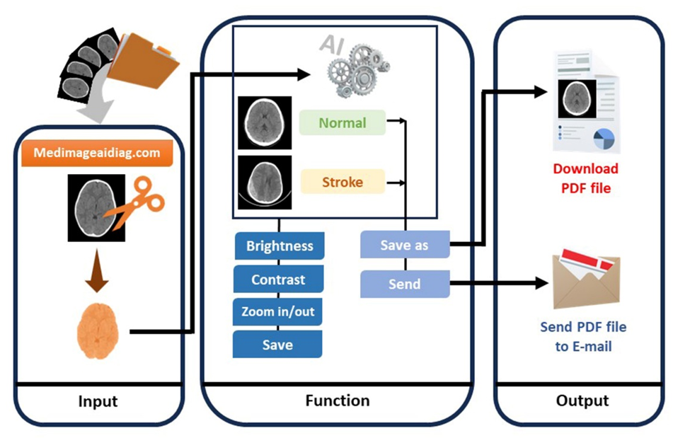
บทนำ: โรคหลอดเลือดสมองมีความเสี่ยงสูงในผู้สูงวัย การวินิจฉัยด้วยภาพถ่ายเอกซเรย์คอมพิวเตอร์มีความต้องการความแม่นยำ ความเชี่ยวชาญในการแปลผลเป็นสิ่งจำเป็น และปัญญาประดิษฐ์เข้ามามีส่วนช่วยให้รังสีแพทย์แปลผลถูกต้องมากขึ้นในปัจจุบัน วัตถุประสงค์: งานวิจัยนี้มีวัตถุประสงค์เพื่อพัฒนาเว็บแอปพลิเคชันสำหรับตรวจหาโรคหลอดเลือดสมองในภาพเอกซเรย์คอมพิวเตอร์สมองโดยอาศัยปัญญาประดิษฐ์ในการแยกภาพโรคหลอดเลือดสมอง วิธีการศึกษา: ข้อมูลภาพเอกซเรย์คอมพิวเตอร์สมองจำนวน 1,636 ภาพ (1,111 ภาพปกติ และ 525 ภาพโรคหลอดเลือดสมอง) ถูกใช้ในงานวิจัยนี้ ภาพประมาณร้อยละ 70 (1,175 ภาพ) ของภาพทั้งหมดถูกใช้ในการฝึกฝนโมเดล ร้อยละ 20 (329 ภาพ) ใช้ในการตรวจสอบ และร้อยละ 10 (132 ภาพ) ใช้ในการทดสอบปัญญาประดิษฐ์โดยใช้โมเดลการเรียนรู้เชิงลึกวีจีจี-16 ค่าความถูกต้อง ความไว ความจำเพาะ เอฟวัน-สกอร์ และพื้นที่ใต้กราฟ ถูกใช้ในประเมินประสิทธิภาพของเว็บแอปพลิเคชัน และทำการประเมินการออกแบบและการใช้งานเว็บแอปพลิเคชันด้วยการให้คะแนน 5 ระดับ โดยรังสีแพทย์ 3 ท่าน ผลการศึกษา: ผลการวิจัยพบว่าค่าความถูกต้อง ความไว ความจำเพาะ เอฟวัน-สกอร์ และพื้นที่ใต้กราฟ มีค่าเท่ากับ 0.969 0.952 0.978 0.952 และ 0.965 ตามลำดับ คะแนนการออกแบบและการใช้งานประเมินโดยรังสีแพทย์มีค่าเท่ากับ 4.13±0.38 และ 4.37±0.33 ตามลำดับ สรุปผลการศึกษา: เว็บแอปพลิเคชันที่พัฒนาขึ้นมีประสิทธิภาพในการวินิจฉัยโรคหลอดเลือดสมองในภาพเอกซเรย์คอมพิวเตอร์สมองและง่ายต่อการใช้งานบนระบบอินเตอร์เน็ต
Downloads
เอกสารอ้างอิง
World Health Organization (WHO). World stroke day 2022 [cited 2022 November 17]. Available from: https://www.who.int/srilanka/news/detail/29-10-2022-world-stroke-day-2022
The Ministry of Public Health of Thailand, Public Health Statistics A.D. 2022. [cited 2022 May 14]. Available from: https://spd.moph.go.th/wp-content/uploads/2023/11/Hstatistic65.pdf
González RG. Current State of Acute Stroke Imaging. Stroke. 2013; 44(11):3260-3264 https://doi.org/10.1161/STROKEAHA.113.003229
Birenbaum D, Bancroft LW, Felsberg GJ. Imaging in Acute Stroke. West J Emerg Med. 2011;12(1):67-76.
Guberina N, Dietrich U, Radbruch A, et al. Detection of early infarction signs with machine learning based diagnosis by means of the Alberta Stroke Program Early CT score (ASPECTS) in the clinical routine. Neuroradiology. 2018; 60:889–901.
Kuang H, Najm M, Chakraborty D, et al. Automated ASPECTS on noncontrast CT scans in patients with acute ischemic stroke using machine learning. American Journal of Neuroradiology 2019; 40:33–38
Shafaat O, Bernstock DJ, Shafaat A, et.al. Leveraging artificial intelligence in ischemic stroke imaging. Journal of Neuroradiology. 2022; 49; 343–351.
Pandey N. Lung segmentation from Chest X-Ray dataset. Kaggle Inc [cited 2022 April 28]. Available from: https://www.kaggle.com/code/nikhilpandey
/lung-segmentation-from-chest-x-ray-dataset/
notebook
Tasnia N. brain-stroke-prediction-ct-scan-image-dataset. [cited 2022 May 1]. Available from: https://www.kaggle.com/datasets/noshintasnia/brain-stroke-prediction-ct-scan-image-dataset
Vbookshelf. Brain CT Images with Intracranial Hemorrhage Masks. [cited 2022 May 1]. Available from: https://www.kaggle.com/datasets/vbookshelf/
computed-tomography-ct-images
Kaewlek T, Sitinwan K, Lueangaroon K, Sansuriyawong W. Comparative analysis of deep learning techniques for accurate stroke detection. Journal of Associated Medical Sciences. 2024; 57(2), 49–55.
Varshney P. VGGNet-16 Architecture: A Complete Guide. [cited 2022 January 13]. Available from: https://www.kaggle.com/code/blurredmachine/vggnet-16-architecture-a-complete-guide
Vogt WP. Dictionary of statistics and methodology. 1999, 2nd ed, Sage: Thousand Oaks, California.
Joshi A, Kale S, Chandel S, Pal DK. Likert Scale: Explored and Explained. Current Journal of Applied Science and Technology, 2015; 7(4), 396–403.
Mcleod S. Likert Scale Questionnaire: Examples & Analysis. [cited 2023 August 13]. Available from: https://www.simplypsychology.org/likert-scale.html
Chen YT, Chen YL, Chen YY, Huang YT, Wong HF, Yan JL, et al. Deep Learning–Based Brain Computed Tomography Image Classification with Hyperparameter Optimization through Transfer Learning for Stroke. Diagnostics 2022; 12, 807.
Ozaltin O, Coskun O, Yeniay O, Subasi A. A Deep Learning Approach for Detecting Stroke from Brain CT Images Using OzNet. Bioengineering 2022; 9: 783.
Li S, Zheng J, Li D. Precise segmentation of non-enhanced computed tomography in patients with ischemic stroke based on multi-scale U-Net deep network model. Computer methods and programs in biomedicine. 2021; 208:106278.
Guerrero R, Qin C, Oktay O, Bowles C, Chen L, Joules R, et al. White matter hyperintensity and stroke lesion segmentation and differentiation using convolutional neural networks. Neuroimage Clin. 2018; 17:918-34.
Phong TD, Duong HN, Nguyen HT, Trong NT, Nguyen VH, Hoa TV, et al. Brain hemorrhage diagnosis by using deep learning. Proceedings of the 2017 International Conference on Machine learning and soft Computing; Ho Chi Minh City, Vietnam. Association for Computing Machinery; 2017. p. 34–9.
Gaidhani BR, Rajamenakshi RR, Sonavane S. Brain stroke detection using convolutional neural network and deep learning models. 2019, 2nd international conference on intelligent communication and computational techniques (ICCT); 2019; Sept 28-29. Jaipur, India. 2019.
Mouridsen K, Thurner P, Zaharchuk G. Artificial intelligence applications in stroke. Stroke. 2020; 51(8):2573-9.
Ding L, Liu C, Li Z, Wang Y. Incorporating artificial intelligence into stroke care and research. Stroke. 2020; 51(12): e351-e4.
Ammar M, Lamria MA, Mahmoudib S, Laidia A. Deep Learning Models for Intracranial Hemorrhage Recognition: A comparative study. Procedia Computer Science. 2022;196: 418–425.
Vamsi B, Bhattacharyya D, Midhunchakkravarthy D, Kim JY. Early Detection of Hemorrhagic Stroke Using a Lightweight Deep Learning Neural Network Model. Traitement du signal. 2021;38(6): 1727-1736.
Soun JE, Chow DS, Nagamine M, Takhtawala RS, Filippi CG, Yu W, et al. Artificial intelligence and acute stroke imaging. AJNR Am J Neuroradiol. 2021; 42(1): 2-11.
ดาวน์โหลด
เผยแพร่แล้ว
รูปแบบการอ้างอิง
ฉบับ
ประเภทบทความ
สัญญาอนุญาต
ลิขสิทธิ์ (c) 2024 สมาคมรังสีเทคนิคแห่งประเทศไทย

อนุญาตภายใต้เงื่อนไข Creative Commons Attribution-NonCommercial-NoDerivatives 4.0 International License.
บทความที่ได้รับการตีพิมพ์เป็นลิขสิทธิ์ของสมาคมรังสีเทคนิคแห่งประเทศไทย (The Thai Society of Radiological Technologists)
ข้อความที่ปรากฏในบทความแต่ละเรื่องในวารสารวิชาการเล่มนี้เป็นความคิดเห็นส่วนตัวของผู้เขียนแต่ละท่านไม่เกี่ยวข้องกับสมาคมรังสีเทคนิคแห่งประเทศไทยและบุคคลากรท่านอื่น ๆในสมาคม ฯ แต่อย่างใด ความรับผิดชอบองค์ประกอบทั้งหมดของบทความแต่ละเรื่องเป็นของผู้เขียนแต่ละท่าน หากมีความผิดพลาดใดๆ ผู้เขียนแต่ละท่านจะรับผิดชอบบทความของตนเองแต่ผู้เดียว


