
การวิเคราะห์พื้นผิวโดยใช้เมทริกซ์การเกิดร่วมระดับสีเทาสำหรับการประเมินคุณภาพของภาพสมองระหว่างเครื่องเอกซเรย์คอมพิวเตอร์สองรุ่น
คำสำคัญ:
เอกซเรย์คอมพิวเตอร์สมอง, การวิเคราะห์พื้นผิว, ค่าปริมาณรังสีวินิจฉัยอ้างอิง, เมทริกซ์การเกิดร่วมระดับสีเทา, คุณภาพของภาพบทคัดย่อ
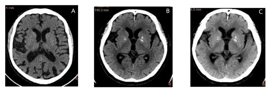
บทนำ: เทคนิคเมทริกซ์การเกิดร่วมระดับสีเทาเป็นวิธีการวิเคราะห์พื้นผิวที่ได้รับความนิยมอย่างมากในปัจจุบันในการใช้ประเมินคุณภาพภาพซีทีในเชิงตัวเลข วัตถุประสงค์: การศึกษานี้มีวัตถุประสงค์เพื่อประเมินคุณภาพของภาพเอกซเรย์คอมพิวเตอร์ส่วนสมองที่ได้จากเครื่องเอกซเรย์คอมพิวเตอร์แบบหลายหัววัดสองรุ่นจากผู้ผลิตที่แตกต่างกัน โดยใช้การวิเคราะห์พื้นผิวเชิงตัวเลขเพื่อกำหนดค่ากระแสไฟฟ้าหลอด (มิลลิแอมป์) ที่เหมาะสมที่สุดของเครื่องทดลองให้มีคุณภาพภาพเทียบเท่าเครื่องมาตรฐาน วิธีการศึกษา: เป็นการวิจัยเชิงวิเคราะห์โดยเก็บข้อมูลย้อนหลังของผู้ป่วยจำนวน 146 ราย เปรียบเทียบภาพที่ได้จากเครื่องมาตรฐาน (120 กิโลโวลต์, 300 มิลลิแอมป์, ไม่ใช้เทคนิคลดปริมาณรังสี) กับภาพจากเครื่องทดลอง (120 กิโลโวลต์, ปรับ มิลลิแอมป์ เป็น 280, 300, 310, และ 315, สร้างภาพด้วยเทคนิค IR-B) ทำการวิเคราะห์พื้นผิวด้วยเมทริกซ์การเกิดร่วมระดับสีเทา (GLCM) จากบริเวณที่สนใจ (ROI) ที่ฐานกะโหลกศีรษะ ก้านสมอง และสมองกลีบข้าง จากนั้นคำนวณหาค่าเฉลี่ย ค่าความเปรียบต่าง ค่าความเป็นเนื้อเดียวกัน และค่าเอนโทรปี เพื่อเปรียบเทียบเชิงปริมาณ ผลการศึกษา: ค่าการวิเคราะห์พื้นผิวที่คำนวณได้ ได้แก่ ค่าระดับสีเทาเฉลี่ย (66.24–183.25), ค่าความเปรียบต่าง (53.17–93.25), ค่าความเป็นเนื้อเดียวกัน (0.14–0.24), และค่าเอนโทรปี (4.24–6.48) พบว่าภาพที่ได้จากเครื่องทดลองที่ตั้งค่า 120 กิโลโวลต์ และ 310 มิลลิแอมป์ ให้ค่าการวิเคราะห์พื้นผิวใกล้เคียงกับภาพมาตรฐานมากที่สุด สรุปผลการศึกษา: คุณภาพของภาพเอกซเรย์คอมพิวเตอร์จากเครื่องทดลองที่ปรับค่ากระแสไฟฟ้าหลอดเป็น 310 มิลลิแอมป์ โดยใช้เทคนิค IR-B) มีคุณภาพใกล้เคียงกับเครื่องมาตรฐาน (300 มิลลิแอมป์) โดยไม่ปรับค่ากิโลโวลต์ ผลการศึกษานี้ยืนยันการตั้งค่ากระแสหลอดเอกซเรย์ที่เหมาะสมที่สุดเพื่อคงไว้ซึ่งคุณภาพของภาพที่เทียบเท่ากัน ซึ่งสามารถนำไปใช้ในการกำหนดค่ากลางสำหรับมาตรฐานการประกันคุณภาพ ของภาพในเครือข่ายโรงพยาบาล
Downloads
เอกสารอ้างอิง
Dieckmeyer M, Sollmann N, Kupfer K, et al. Computed tomography of the head. Radiology. 2023;308(1):e221687. doi:10.1148/radiol.221687.
American College of Radiology. Radiation dose from X-ray and CT exams. RadiologyInfo.org. Updated 2018 Aug 3. Accessed Aug 20, 2025.
Saengphet S, Pintavirooj C. Screening of ischemic stroke in CT brain using image segmentation and texture analysis. In: Proceedings of the 7th International Conference on Engineering, Applied Sciences and Technology (ICEAST); 1–3 April 2021; Pattaya, Thailand.
Singh S, Kalra MK, Gilman MD, et al. Innovations in CT dose reduction strategy: application of adaptive statistical iterative reconstruction algorithm. AJR Am J Roentgenol. 2011;197(5):W833–W839.
Baskan O, Alagoz E, Gunes C, et al. Effect of radiation dose reduction on image quality in adult head CT. J Appl Clin Med Phys. 2015;16(3):287–299.
Philips Healthcare. Ingenuity CT family: iDose4 and O-MAR technologies. Published 2017.
GE HealthCare. Optima CT660: ASiR inside—A leap ahead in dose management.
Castellano G, Bonilha L, Li LM, Cendes F. Texture analysis of medical images. Clin Radiol. 2004;59(12):1061–1069.
Skogen K, Ganeshan B, Good C, Critchley G, Miles K. Measurements of heterogeneity in gliomas on computed tomography: relationship to tumor grade. J Neurooncol. 2013;111(2):213–219.
Ali R, Menaka R. Ischemic stroke lesion detection, characterization and classification in CT images with optimal feature selection.Biomed Eng Lett. 2020;10(3):333–344. doi:10.1007/s13534-020-00163-1.
Lambin P, Leijenaar RTH, Deist TM, et al. CT texture analysis challenges: influence of acquisition and reconstruction parameters. Diagnostics (Basel). 2020;10(5):258.
Lubner MG, El-Haddad G, El-Haddad M, et al. Quantitative assessment of variation in CT parameters on texture features. AJNR Am J Neuroradiol. 2017;38(5):981–986.
Varini M, Carbone SF, Ropolo M, et al. Physical image quality of different scanners for head CT imaging: a phantom study. Phys Med. 2021;79:15–28.
Yang Y, Liu W, Kim K, et al. Fully automated image quality evaluation on patient CT: multi-vendor and multi-reconstruction study. PLoS One. 2022;17(7):e0271724.
Iamsuk T, Meekingthong C, Prasertsilpakul W, Sodkokkruad P, Asavaphatiboon S. Comparison between conventional pre-contrast and virtual non-contrast images from IQon spectral CT. Thai J Rad Tech. 2022;47(1):83–92.
Pimsorn P. Factors affecting size-specific dose estimates in computed tomography using automatic tube current modulation. Thai J Rad Tech. 2022;47(1):43–54.
Chokchai B. Assessment of radiation dose from abdominal computed tomography at Maharat Nakhon Ratchasima Hospital. Thai J Rad Tech. 2023;48(1):110–118.
Wattanasriroj Y, Punthaisiri P, Kingkaew S, Wisetrinthong M, Oonsiri S. The effect of tube voltage and current on the CT number and relative electron density in computed tomography simulator. Thai J Rad Tech. 2023;48(1):18–28.
Admontree S, Asavaphatiboon S. Management of appropriate radiation dose in pediatric patients from computed tomography scans using diagnostic reference levels (DRLs). Thai J Rad Tech. 2024;49(1):129-41.
Singh S, Kalra MK, Gilman MD, et al. Adaptive statistical iterative reconstruction technique for radiation dose reduction in CT: a systematic review. Radiology. 2020;295(3):671-684.doi:10.1148/radiol.2020192258
Bodelle B, Fischbach F, Klotz E, et al. Low-dose cerebral CT using iterative reconstruction: image quality and diagnostic confidence.
Eur Radiol. 2020;30(6):3307-3316. doi:10.1007/s00330-020-06707-8
Mackin D, Fave X, Zhang L, et al. Measuring computed tomography scanner variability of radiomics features. Invest Radiol. 2020;55(12):747-754. doi:10.1097/RLI.0000000000000699
American College of Radiology. ACR–SPR practice parameter for diagnostic reference levels and achievable doses in medical X-ray imaging. Revised 2021.
ดาวน์โหลด
เผยแพร่แล้ว
รูปแบบการอ้างอิง
ฉบับ
ประเภทบทความ
สัญญาอนุญาต
ลิขสิทธิ์ (c) 2025 สมาคมรังสีเทคนิคแห่งประเทศไทย

อนุญาตภายใต้เงื่อนไข Creative Commons Attribution-NonCommercial-NoDerivatives 4.0 International License.
บทความที่ได้รับการตีพิมพ์เป็นลิขสิทธิ์ของสมาคมรังสีเทคนิคแห่งประเทศไทย (The Thai Society of Radiological Technologists)
ข้อความที่ปรากฏในบทความแต่ละเรื่องในวารสารวิชาการเล่มนี้เป็นความคิดเห็นส่วนตัวของผู้เขียนแต่ละท่านไม่เกี่ยวข้องกับสมาคมรังสีเทคนิคแห่งประเทศไทยและบุคคลากรท่านอื่น ๆในสมาคม ฯ แต่อย่างใด ความรับผิดชอบองค์ประกอบทั้งหมดของบทความแต่ละเรื่องเป็นของผู้เขียนแต่ละท่าน หากมีความผิดพลาดใดๆ ผู้เขียนแต่ละท่านจะรับผิดชอบบทความของตนเองแต่ผู้เดียว


