Sero-prevalence and PCR identification of lumpy skin disease virus in cattle at Mymensingh district of Bangladesh https://doi.org/10.12982/VIS.2025.038
Main Article Content
Abstract
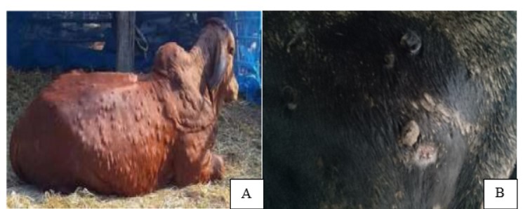
Lumpy skin disease (LSD) is a transboundary, an infectious viral disease of cattle causing substantial economic losses due to skin damage and decreased animal production. This study aimed to determine the serological prevalence and molecular detection of lumpy skin disease virus (LSDV) from cattle in Mymensingh district, Bangladesh with assessment of risk factors. A total of 200 samples consists of whole blood (184), tissue (10) and pus (6) were collected from three different Upazilas of Mymensingh and tested using indirect enzyme linked immunosorbent assay (ELISA) kit. The DNA was extracted from whole blood using a DNA extraction kit and kept at -20°C until further analysis. Extracted DNA was used in a PCR test to detect lumpy skin disease virus (LSDV). Overall, the sero-prevalence of LSD among cattle in Mymensingh was 28.26% (52/184) (CI: 19.36-38.61). Fulbaria Upazilas showed the highest prevalence of 30% (24/80) compared to Mymensingh Sadar 24% (12/50) and Muktagacha 29.63% (16/54). Moreover, the risk of getting LSD infection was lower among cross breed (OR=0.27, CI: -0.1-0.75) and female animals (OR=0.39, CI: -0.15-1.03). Furthermore, freely grazing animal 29.17% (CI: 12.61-51.09) and young group 72.73% (CI: 39.03-93.98) of animal showed risk in having lumpy skin disease. LSD virus was detected in the sero-positive blood sample (100%), tissue sample (90%), and pus/edema fluid discovered (83.33%) after amplification of a 192 bp DNA fragment. The findings of this study will be helpful in creating efficient methods for identifying and managing LSD in Bangladesh and avoiding the financial losses connected with dairy production.
Article Details

This work is licensed under a Creative Commons Attribution 4.0 International License.
Publishing an article with open access in Veterinary Integrative Sciences leaves the copyright with the author. The article is published under the Creative Commons Attribution License 4.0 (CC-BY 4.0), which allows users to read, copy, distribute and make derivative works from the material, as long as the author of the original work is cited.
References
Abera, Z., Degefu, H., Gari, G., Kidane, M., 2015. Sero-prevalence of lumpy skin disease in selected districts of West Wollega zone, Ethiopia. BMC Vet. Res. 17(11), 135.
Babiuk, S., Bowden, T.R., Parkyn, G., Dalman, B., Manning, L., Neufeld, J., Embury-Hyatt, C., Copps, J., Boyle, D.B., 2008. Quantification of lumpy skin disease virus following experimental infection in cattle. Transbound. Emerg. Dis. 55(7), 299–307.
Badhy, S.C., Chowdhury, M.G.A., Settypalli, T.B.K., Cattoli, G., Lamien, C.E., Fakir, M. A.U., Akter, S., Osmani, M.G., Talukdar, F., Begum, N., Khan, I.A., Rashid, M.B., Sadekuzzaman, M., 2021. Molecular characterization of lumpy skin disease virus (LSDV) emerged in Bangladesh reveals unique genetic features compared to contemporary field strains. BMC Vet. Res. 17(61), 1-11.
Chihota, C.M., Rennie, L.F., Kitching, R.P., Mellor, P.S., 2003. Attempted mechanical transmission of lumpy skin disease virus by biting insects. Med. Vet. Entomol. 17(3), 294–300.
Das, M., Chowdhury, M.S.R., Akter, S., Mondal, A.K., Uddin, M.J., Rahman, M.M., Rahman, M.M., 2021. An updated review on lumpy skin disease: perspective of Southeast Asian countries. J. Adv. Biotechnol. Exp. Ther. 4(3), 322–333.
DLS, 2019. Situation Report: Lumpy Skin Disease in Bangladesh Background. Available online: https://www.oie.int/fileadmin/Home/eng/Animal_Health_in_the_World/docs /pdf/Disease_cards/lumpy_skin_disease_2019_final.pdf.
Dubie, T., Hussen, A.F., Dereje, B., Negash, W., Hamid, M., 2022. Seroprevalence and associated risk factors of lumpy skin disease of cattle in selected districts of Afar Region, Ethiopia. Vet. Med. Res. Rep. 16(13), 191–199.
Gari, G., Grosbois, V., Waret-Szkuta, A., Babiuk, S., Jacquiet, P., Roger, F., 2012. Lumpy skin disease in Ethiopia: Seroprevalence study across different agro-climate zones. Acta Tropica. 123(2), 101–106.
Giasuddin, M., Yousuf, M., Hasan, M., Rahman, M., Hassan, M., Ali, M., 2020. Isolation and molecular identification of Lumpy Skin Disease (LSD) virus from infected cattle in Bangladesh. Bangladesh. J. Livest. Res. 26(1–2), 15–20.
Haegeman, A., De Leeuw, I., Mostin, L., Van Campe, W., Aerts, L., Vastag, M., De Clercq, K., 2020. An Immunoperoxidase Monolayer Assay (IPMA) for the detection of lumpy skin disease antibodies. J. Virol. Methods. 277, 113800.
Ireland, D.C., Binepal, Y.S., 1998. Improveddetection of capripoxvirus in biopsy samples byPCR. J. Virol. Methods. 74 (1), 1–7.
Irons, P.C., Tuppurainen, E.S.M., Venter, E.H., 2005. Excretion of lumpy skin disease virus in bull semen. Theriogenology. 63(5), 1290–1297.
Khan, A., Du, X., Hussain, R., Kwon, O.D., 2022. Lumpy skin disease: a threat to the livestock industry - a review. Agrobiol. Rec. 9, 22–36.
Kitching, R.P., Taylor, W.P., 1985. Transmission of capripoxvirus. Res. Vet. Sci. 39(2), 196–199.
Molla, W., Frankena, K., Gari, G., Kidane, M., Shegu, D., de Jong, M.C.M., 2018. Seroprevalence and risk factors of lumpy skin disease in Ethiopia. Prev. Vet. Med. 160, 99–104.
Osuagwuh, U.I., Bagla, V., Venter, E.H., Annandale, C.H., Irons, P.C., 2007. Absence of lumpy skin disease virus in semen of vaccinated bulls following vaccination and subsequent experimental infection. Vaccine. 25(12), 2238–2243.
Quinn, P.J., Markey, B.K., Carter, M.E., Donnelly, W.J., Leonard, F.C., 2002. Veterinary Microbiology and Microbial Disease. Iowa State University Press, Ames, Iowa, USA, pp. 536.
Radostits, O.M., Gay, C.C., Hinchcliff, K.W., Constable, P.D., 2007 Veterinary medicine -a textbook of the diseases of cattle, horses, sheep, pigs and goats, 10th edition. Saunders, USA, pp. 2065.
Sergeant ESG, 2018. Epitools Epidemiological Calculators. Available online: https://epitools.ausvet.com.au/.
Tuppurainen, E.S.M., Oura, C.A.L., 2012. Review: lumpy skin disease: an emerging threat to Europe, the Middle East and Asia. Transbound. Emerg. Dis. 59(1), 40–48.
Tuppurainen, E.S.M., Stoltsz, W.H., Troskie, M., Wallace, D.B., Oura, C.A.L., Mellor, P.S., Coetzer, J.A.W., Venter, E.H., 2011. A potential role for ixodid (hard) tick vectors in the transmission of lumpy skin disease virus in cattle. Transbound. Emerg. Dis. 58(2), 93–104.
WOAH, 2021. Lumpy skin disease. Available online: http://www.woah.org/app/uploads /2021/03/lumpy-skin-disease.pdf.

