Fatal vascular pythiosis in a dog from Thailand: Clinical presentation and pathological findings—first report: A case report https://doi.org/10.12982/VIS.2026.069
Article Sidebar
Main Article Content
Abstract
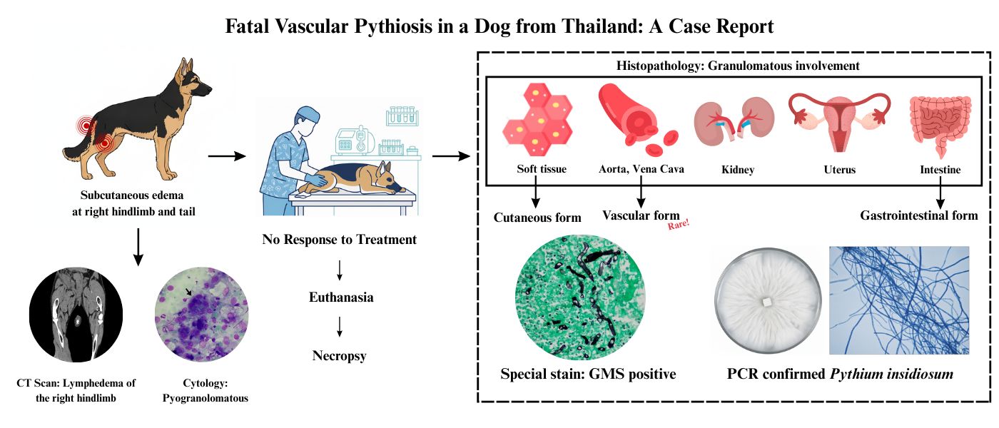
Vascular involvement in Pythium insidiosum infection is exceedingly rare in dogs and often results in delayed diagnosis due to nonspecific clinical signs. A one-year-old female German Shepherd presented with subcutaneous edema and mass-like lesions on the right hindlimb, caudal abdomen, and tail base. Computed tomography lymphangiography revealed lymphedema with impaired lymphatic drainage, and cytologic evaluation demonstrated pyogranulomatous inflammation, including multinucleated giant cells containing poorly stained, contorted fungal hyphae within the cytoplasm. Despite right hindlimb amputation, the disease progressed to ischemic necrosis, gangrene and eventual autoamputation of the left hindlimb and tail. Severe P. insidiosum infection was confirmed by histopathologic examination and immunochromatographic testing. Clinical deterioration continued despite aggressive antifungal and antimicrobial therapy, leading to euthanasia. Postmortem examination revealed extensive pyogranulomatous and necrotizing inflammation involving the aorta, caudal vena cava, kidneys, uterus and intestines. Broad, ribbon-like, sparsely septate hyphae were observed and confirmed as P. insidiosum by culture, and by Internal Transcribed Spacer Polymerase Chain Reaction. This case represents the first reported instance of vascular pythiosis in a dog, highlighting its rarity, severity and poor prognosis. Early recognition of vascular involvement is critical, as delayed diagnosis and limited therapeutic response are frequently fatal
Article Details

This work is licensed under a Creative Commons Attribution 4.0 International License.
Publishing an article with open access in Veterinary Integrative Sciences leaves the copyright with the author. The article is published under the Creative Commons Attribution License 4.0 (CC-BY 4.0), which allows users to read, copy, distribute and make derivative works from the material, as long as the author of the original work is cited.
References
Camus, A.C., Grooters, A.M., Aquilar, R.F., 2004. Granulomatous Pneumonia Caused by Pythium Insidiosum in a Central American Jaguar, Panthera Onca. J. Vet. Diagn. Invest. 16(6), 567–571.
Chindamporn, A., Kammarnjessadakul, P., Kesdangsakonwut, S., Banlunara, W., 2020. A case of canine cutaneous pythiosis in Thailand. Access Microbiol. 2(4), e000109.
Chitasombat, M.N., Larbcharoensub, N., Chindamporn, A., Krajaejun, T., 2018. Clinicopathological features and outcomes of pythiosis. Int. J. Infect. Dis. 71, 33–41.
Chitasombat, M.N., Petchkum, P., Horsirimanont, S., Sornmayura, P., Chindamporn, A., Krajaejun, T., 2018. Vascular pythiosis of the carotid artery with meningitis and cerebral septic emboli: a case report and literature review. Med. Mycol. Case Rep. 21, 57–62.
Davis, D.J., Lanter, K., Makselan, S., Bonati, C., Asbrock, P., Ravishankar, J.P., et al., 2006. Relationship between temperature optima and secreted protease activities of three Pythium species and pathogenicity toward plant and animal hosts. Mycol. Res. 110(1), 96–103.
De Cock, A.W., Mendoza, L., Padhye, A.A., Ajello, L., Kaufman, L., 1987. Pythium insidiosum sp. nov., the etiologic agent of pythiosis. J. Clin. Microbiol. 25(2), 344–349.
Elshafie, N.O., Hanlon, J., Malkawi, M., Sayedahmed, E.E., Guptill, L.F., Jones-Hall, Y.L., Santos, A.P., 2022. Nested PCR detection of Pythium sp. from formalin-fixed, paraffin-embedded canine tissue sections. Vet. Sci. 9(8), 444.
Gaastra, W., Lipman, L.J.A., De Cock, A.W.A.M., Exel, T.K., Pegge, R.B.G., Scheurwater, J., et al., 2010. Pythium insidiosum: An overview. Vet. Microbiol. 146(1–2), 1–16.
Grooters, A.M., 2022. Pythiosis, lagenidiosis, paralagenidiosis, entomophthoromycosis, and mucormycosis. In: Greene, C.E. (Ed.), Greene’s infectious diseases of the dog and cat, (5th edition). Elsevier, New York, pp. 1105–1112.
Imwidthaya, P., 1994. Human pythiosis in Thailand. Postgrad. Med. J. 70(826), 558–560.
Keoprasom, N., Chularojanamontri, L., Chayakulkeeree, M., Chaiprasert, A., Wanachiwanawin, W., Ruangsetakit, C., 2013. Vascular pythiosis in a thalassemic patient presenting as bilateral leg ulcers. Med. Mycol. Case Rep. 2, 25–28.
Khunkhet, S., Rattanakaemakorn, P., Rajatanavin, N., 2015. Pythiosis presenting with digital gangrene and subcutaneous nodules mimicking medium vessel vasculitis. JAAD Case Rep. 1(6), 399–402.
Krajaejun, T., Sathapatayavongs, B., Pracharktam, R., Nitiyanant, P., Leelachaikul, P., Wanachiwanawin, W., Chaiprasert, A., Assanasen, P., Saipetch, M., Mootsikapun, P., Chetchotisakd, P., Lekhakula, A., Mitarnun, W., Kalnauwakul, S., Supparatpinyo, K., Chaiwarith, R., Chiewchanvit, S., Tananuvat, N., Srisiri, S., Suankratay, C., Kulwichit, W., Wongsaisuwan, M., Somkaew, S., 2006. Clinical and epidemiological analyses of human pythiosis in Thailand. Clin. Infect. Dis. 43(5), 569–576.
MacDonald, E., Millward, L., Ravishankar, J.P., Money, N.P., 2002. Biomechanical interaction between hyphae of two Pythium species (Oomycota) and host tissues. Fungal Genet. Biol. 37(3), 245–249.
Norasetthada, A., 2025. Vasculitis mimics caused by Pythium infection and literature review. Thai J. Rheu. 2(2), 27–36.
Pan, J.H., Kerkar, S.P., Siegenthaler, M.P., Hughes, M., Pandalai, P.K., 2014. A complicated case of vascular Pythium insidiosum infection treated with limb-sparing surgery. Int. J. Surg. Case Rep. 5(10), 677–680.
Peano, A., Min, A.R.M., Fondati, A., Romano, E., Brachelente, C., Porcellato, I., Amore, A., Pasquetti, M., 2023. Cutaneous pythiosis in 2 dogs, Italy. Emerg. Infect. Dis. 29(7), 1447–1450.
Ravishankar, J.P., Davis, C.M., Davis, D.J., MacDonald, E., Makselan, S.D., Millward, L., Money, N.P., 2001. Mechanics of solid tissue invasion by the mammalian pathogen Pythium insidiosum. Fungal Genet. Biol. 34(3), 167–175.
Rivierre, C., Laprie, C., Guiard-Marigny, O., Bergeaud, P., Berthelemy, M., Guillot, J., 2005. Pythiosis in Africa. Emerg. Infect. Dis. 11(3), 479–481.
Rotchanapreeda, T., Sae-Chew, P., Lohnoo, T., Yingyong, W., Rujirawat, T., Kumsang, Y., Payattikul, P., Jaturapaktrarak, C., Intaramat, A., Pathomsakulwong, W., Yurayart, C., Krajaejun, T., 2021. Immunological cross-reactivity of proteins extracted from the oomycete Pythium insidiosum and the fungus Basidiobolus ranarum compromises the detection specificity of immunodiagnostic assays for pythiosis. J. Fungi. 7(6), 474.
Salipante, S.J., Hoogestraat, D.R., SenGupta, D.J., Murphey, D., Panayides, K., Hamilton, E., Castañeda-Sánchez, I., Kennedy, J., Monsaas, P.W., Mendoza, L., Stephens, K., Dunn, J.J., Cookson, B.T., 2012. Molecular diagnosis of subcutaneous Pythium insidiosum infection by use of PCR screening and DNA sequencing. J. Clin. Microbiol. 50(4), 1480–1483.
Samanta, I., 2015. Cutaneous, subcutaneous and systemic mycology. In: Veterinary Mycology. Springer, New Delhi, pp. 127–132.
Schloemer, N.J., Lincoln, A.H., Mikhailov, T.A., Collins, C.L., Di Rocco, J.R., Kehl, S.C., Chusid, M.J., 2013. Fatal disseminated Pythium insidiosum infection in a child with Diamond-Blackfan anemia. Infect. Dis. Clin. Pract. 21(4), e24–e26.
Shipton, W.A., 1987. Pythium destruens sp. nov., an agent of equine pythiosis. J. Med. Vet. Mycol. 25(3), 137–151.
Sudjaritruk, T., Sirisanthana, V., 2011. Successful treatment of a child with vascular pythiosis. BMC Infect. Dis. 11(1), 33.
Sukanan, P., Suparp, B., Yongsiri, S., Chansiripornchai, P., Kesdangsakonwut, S., 2022. Successful management of colonic pythiosis in two dogs in Thailand using antifungal therapy. Vet. Med. Sci. 8(6), 2283–2291.
Thitithanyanont, A., Mendoza, L., Chuansumrit, A., Pracharktam, R., Laothamatas, J., Sathapatayavongs, B., Lolekha, S., Ajello, L., 1998. Use of an immunotherapeutic vaccine to treat a life-threatening human arteritic infection caused by Pythium insidiosum. Clin. Infect. Dis. 27(6), 1394–1400.
Thongsuk, P., Plongla, R., Thammahong, A., Tiewsurin, J., Worasilchai, N., Chindamporn, A., Suankratay, C., 2021. Vascular pythiosis caused by Pythium aphanidermatum: the first case report in Asia. Eur. J. Med. Res. 26, 132.
White, T.J., Bruns, T., Lee, S., Taylor, J., 1990. Amplification and direct sequencing of fungal ribosomal RNA genes for phylogenetics. In: Innis, M.A., Gelfand, D.H., Sninsky, J.J., White, T.J. (Eds.), PCR Protocols: a guide to methods and applications. Academic Press, San Diego, pp. 315–322.
Worasilchai, N., Permpalung, N., Chongsathidkiet, P., Leelahavanichkul, A., Mendoza, A.L., Palaga, T., Reantragoon, R., Finkelman, M., Sutcharitchan, P., Chindamporn, A., 2018. Monitoring anti-Pythium insidiosum IgG antibodies and (1→3)-β-D-glucan in vascular pythiosis. J. Clin. Microbiol. 56(8), e00610-18.
Yolanda, H., Krajaejun, T., 2022. Global distribution and clinical features of pythiosis in humans and animals. J. Fungi. 8(2), 182.
