Digital H&E colorimetric profiling of ovine brain microstructures https://doi.org/10.12982/VIS.2026.080
Article Sidebar
Main Article Content
Abstract
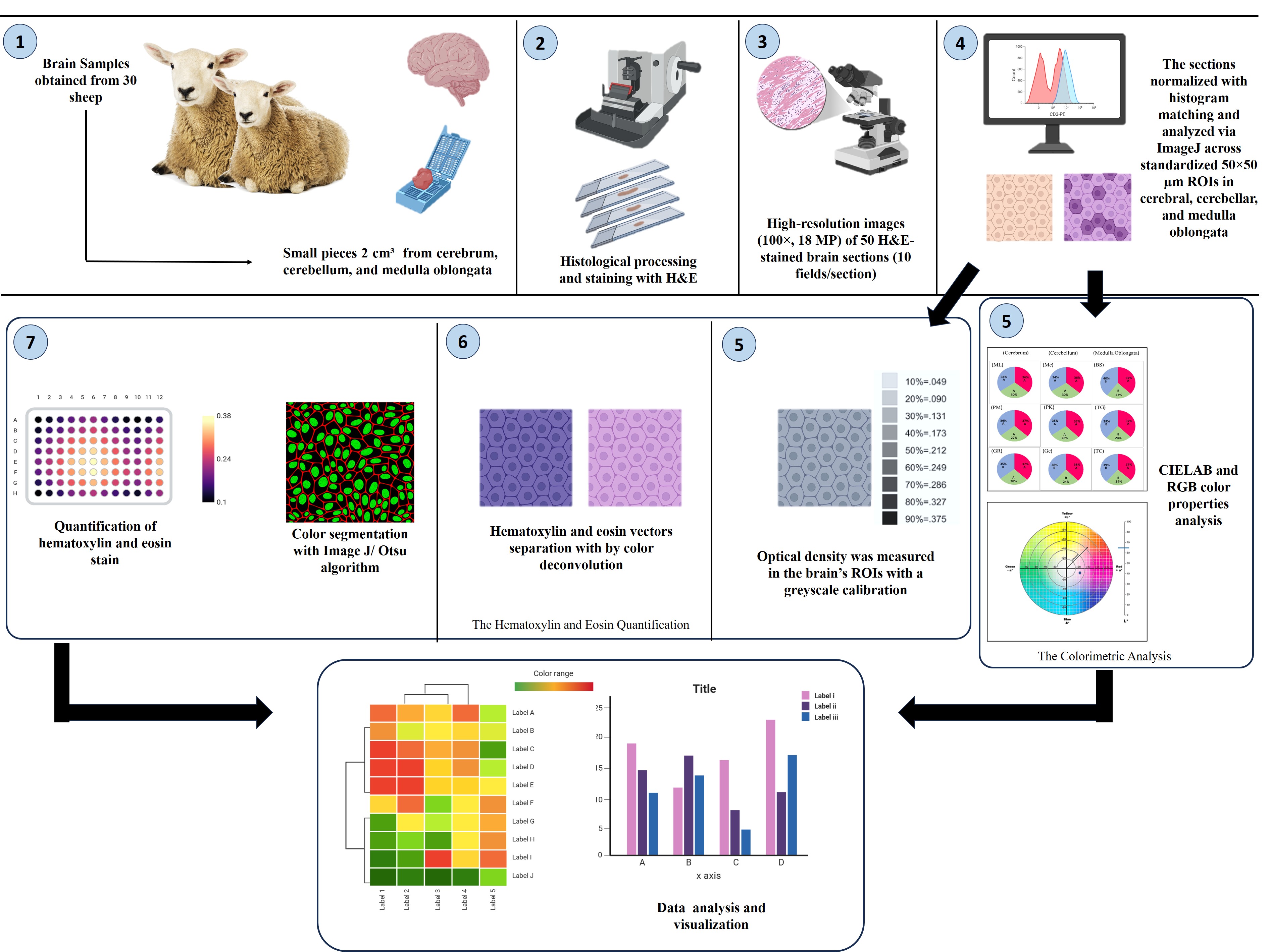
Differences in staining techniques and inconsistencies of staining protocols lead to different visual appearances, which in turn affect color quality in the digital images during brain histopathological analysis, therefore, this study aimed to characterize colorimetric profiles for the cerebrum, cerebellum, and medulla oblongata in the sheep brain by measuring optical density, color space decomposition to analyze stain intensity and proportion using both the RGB and CIELAB color models. All digital images were normalized using a histogram matching algorithm. The result revealed different stain patterns across the brain microstructure. The cerebellar regions showed a higher hematoxylin optical density compared to the cerebrum and medulla oblongata, while eosin intensity and proportion were the highest in the medullary deep layers. The color channels showed that red intensity was highest in the cerebellum and lowest in the medulla. While the blue channel showed the opposite pattern. Digital colorimetry effectively distinguishes healthy neural tissue variation based on H&E staining profiles, serves as a quantitative baseline that may support the investigation of future diagnostic tools for color-dependent brain lesions, such as ischemia, early infarction, or demyelinating lesions.
Article Details

This work is licensed under a Creative Commons Attribution 4.0 International License.
Publishing an article with open access in Veterinary Integrative Sciences leaves the copyright with the author. The article is published under the Creative Commons Attribution License 4.0 (CC-BY 4.0), which allows users to read, copy, distribute and make derivative works from the material, as long as the author of the original work is cited.
References
Altaey, O.Y., Hasan, A.A., Alhaaik, A.G., 2025. Early-life development of spleen in white rabbit (Oryctolagus cuniculus): a morphometric and histochemical analysis. Vet. Integr. Sci. 23(1), 1-13.
Bankhead, P., 2022. Developing image analysis methods for digital pathology, J. Pathol. 257(4), 391-402.
Banstola, A., Reynolds, J. N., 2022. Mapping sheep to human brain: The need for a sheep brain atlas. Front. Vet. Sci. 9, 961413.
Benton, H.M., Butters, M., Brous, M., Bolon, B., Copeland, K., Fortin, J.S., Chlipala, E., 2025. Utilizing image analysis by optical density to evaluate changes in hematoxylin and eosin staining quality after reagent overuse. J. Histopathol. 48(3), 123-134.
Berlanga, M.L., Phan, S., Bushong, E.A., Wu, S., Kwon, O., Phung, B.S., Lamont, S., Terada, M., Tasdizen, T., Martone, M.E., Ellisman, M.H., 2011. Three-dimensional reconstruction of serial mouse brain sections: solution for flattening high-resolution large-scale mosaics. Front. Neuroanat. 5, 17.
Borah, B.J., Tseng, Y.C., Wang, K.C., Wang, H.C., Huang, H.Y., Chang, K., Lin, J.R., Liao, Y.H., Sun, C.K., 2023. Rapid digital pathology of h&e-stained fresh human brain specimens as an alternative to frozen biopsy. Commun. Med (Lond). 3(1), 77.
Cheng, W.C., 2020. Reproducible color gamut of hematoxylin and eosin-stained images in standard color spaces. J. Pathol. Inform. 11(1), 36.
Chlipala, E., Bendzinski, C.M., Chu, K., Johnson, J.I., Brous, M., Copeland, K., Bolon, B., 2020. Optical density-based image analysis method for the evaluation of hematoxylin and eosin staining precision. J. histotechnol. 43(1), 29-37.
Chu, M.L., Ge, X.Y.M., Eastham, J., Nguyen, T., Fuji, R.N., Sullivan, R., Ruderman, D., 2023. Assessment of color reproducibility and mitigation of color variation in whole slide image scanners for toxicologic pathology. Toxicol. Pathol. 51(6), 313-328.
Distante, A., Distante, C., Distante, W., Wheeler, 2020. Handbook of image processing and computer vision. Springer, Gemany.
Dunn, C., Brettle, D., Cockroft, M., Keating, E., Revie, C., Treanor, D., 2024. Quantitative assessment of H&E staining for pathology: development and clinical evaluation of a novel system. Diagn. Pathol. 19(1), 42.
Dunn, C., Brettle, D., Hodgson, C., Hughes, R., Treanor, D., 2025. An international study of stain variability in histopathology using qualitative and quantitative analysis. J. Pathol. Inform. 17, 100423.
Elazab, N., Gab Allah, W., Elmogy, M., 2024. Computer-aided diagnosis system for grading brain tumor using histopathology images based on color and texture features. BMC Med. Imaging. 24(1), 177.
Fanous, M., Caputo, M.P., Lee, Y.J., Rund, L.A., Best-Popescu, C., Kandel, M.E., Johnson, R.W., Das, T., Kuchan, M.J., Popescu, G., 2020. Quantifying myelin content in brain tissue using color Spatial Light Interference Microscopy (cSLIM). PLoS One. 15(11), e0241084.
Gonzalez, R.C., 2009. Digital image processing. Pearson education. Hudson Street, New York.
Grenko, C.M., Viaene, A.N., Nasrallah, M.P., Feldman, M.D., Akbari, H., Bakas, S., 2020. Towards population-based histologic stain normalization of glioblastoma. Brainlesion. 11992, 44–56.
Hasan, A.A., Altaey, O.Y., Hasso, A.A., 2024. Analysis of chukar partridge wing morphology and morphometry and their implications in flight pattern and behavior. Egyptian J. Vet. Sci. 55(7), 1961-1974.
Inoue, T., Yagi, Y., 2020. Color standardization and optimization in whole slide imaging. Clin. Diagn. Pathol. 4(1), 10-15761.
Irshad, H., Veillard, A., Roux, L., Racoceanu, D., 2013. Methods for nuclei detection, segmentation, and classification in digital histopathology: a review—current status and future potential. IEEE Rev. biomed. Eng. 7, 97-114.
Khan, U., Härkönen, J., Friman, M., Hakimnejad, H., Latonen, L., Kuopio, T., Ruusuvuori, P., 2026. Staining normalization in histopathology: Method benchmarking using multicenter dataset. Sci Rep. 16(1), 11097.
Kleczek, P., Jaworek-Korjakowska, J., Gorgon, M., 2020. A novel method for tissue segmentation in high-resolution H&E-stained histopathological whole-slide images. Comput. Med. Imaging Graph. 79, 101686.
Li, X., Plataniotis, K.N., 2015. A complete color normalization approach to histopathology images using color cues computed from saturation-weighted statistics. IEEE Trans. Biomed. Eng. 62(7), 1862-1873.
Madusanka, N., Jayalath, P., Fernando, D., Yasakethu, L., Lee, B.I., 2023. Impact of H&E stain normalization on deep learning models in cancer image classification: performance, complexity, and trade-offs. Cancers. 15(16), 4144.
Michielli, N., Caputo, A., Scotto, M., Mogetta, A., Pennisi, O.A.M., Molinari, F., Balmativola, D., Bosco, M., Gambella, A., Metovic, J., Tota, D., Carpenito, L., Gasparri, P., Salvi, M., 2022. Stain normalization in digital pathology: clinical multi-center evaluation of image quality. J. pathol. Inform. 13, 100145.
Moghadam, A.Z., Azarnoush, H., Seyyedsalehi, S.A., Havaei, M., 2022. Stain transfer using generative adversarial networks and disentangled features. Comput. Biol. Med. 142, 105219.
Muratbekova, M., Toganas, N., Igali, A., Shagyrov, M., Kadyrgali, E., Yerkin, A., Shamoi, P., 2026. Color models in image processing: a review and experimental comparison. Discov. Appl. Sci . 8, 494.
Murray, S.J., Mitchell, N.L., 2022. The translational benefits of sheep as large animal models of human neurological disorders. Front. Vet. Sci. 9, 831838.
Niazi, M.K.K., Parwani, A.V., Gurcan, M.N., 2019. Digital pathology and artificial intelligence. Lancet Oncol. 20(5), e253-e261.
Petrie, A., Watson, P., 2013. Hypothesis tests, the chisquared test: comparing proportions. In Statistics for veterinary and animal science, 3edition. Wiley Blackwell, Eds, USA, pp. 112–126.
Reschke, M., DiRito, J.R., Stern, D., Day, W., Plebanek, N., Harris, M., Hosgood, S.A., Nicholson, M.L., Haakinson, D.J., Zhang, X., Mehal, W.Z., Ouyang, X., Pober, J.S., Saltzman, W.M., Tietjen, G.T., 2022. A digital pathology tool for quantification of color features in histologic specimens. Bioeng. Transl. Med. 7(1), e10242.
Roetzer, T., Leskovar, K., Peter, N., Furtner, J., Muck, M., Augustin, M., Lichtenegger, A., Nowosielski, M., Hainfellner, J.A., Baumann, B., Woehrer, A., 2019. Evaluating cellularity and structural connectivity on whole brain slides using a custom-made digital pathology pipeline. J. Neurosci. Methods. 311, 215-221.
Ruifrok, A.C., Johnston, D.A., 2001. Quantification of histochemical staining by color deconvolution. Anal. Quant. Cytol. Histol. 23(4), 291-299.
Russ, J.C., 2006. The image processing handbook. CRC press, Boca Raton.
Sornying, P., Kaewnoi, D., Keawchana, N., Khumraksa, P., Ruangpoon, S., Ninwat, S., Sukkarun, P., Arnuphapprasert, A., Suyapoh, W., 2026. The hidden burden: morphological, histological, and pathological features of Paradujardinia halicoris in dugongs (Dugong dugon) from the Andaman coast of Thailand: Characterizing the nematode-induced gastrointestinal lesions in a threatened sirenian species. Vet. Integr. Sci. 24(2), 1-16.
Tosta, T.A.A., de Faria, P.R., Neves, L.A., do Nascimento, M.Z., 2019. Computational normalization of H&E-stained histological images: progress, challenges and future potential. Artif. Intell. Med. 95, 118-132.
Wolfe, D., 2019. Tissue processing. In: Suvarna, S.K., Layton, C., Bancroft, J.D. Amsterdam (Eds.), Bancroft’s theory and practice of histological techniques. Elsevier, Netherlands, pp. 83–92.
