
Tinea corporis from microsporum canis: A case report in 2 patients from 1 asymptomatic feline
Keywords:
M. canis, Zoophilic, Zoophilic, Tinea corporis, Tinea corporis, Wood’s lamp, Wood’s lampAbstract
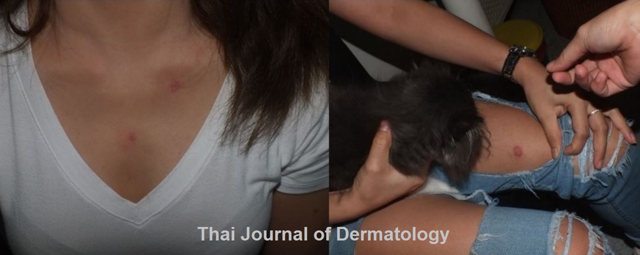
Tinea corporis is a common dermatophyte infection which causes pruritic erythematous scaly papules and plaques. Most common causative organisms are Trichophyton rubrum and Trichophyton mentagrophytes. However, Microsporum canis can be the pathogen leading to the more sudden and extensive clinical manifestations. M. canis is a zoophilic organisms residing in pets such as dogs or cats, whereas the infected pet can be totally asymptomatic. The treatment is usually systemic antifungal for 2-4 weeks.
We report 2 patients presented with pruritic erythematous scaly plaques diagnosed as tinea corporis from Microsporum canis. The patients lived together with their household cat. Examination of the asymptomatic cat, its fur showed positive green color under wood’s lamp and the culture showed Microsporum canis as well. The systemic antifungal was given along with the advice to thoroughly house cleaning and the treatment for the patients’ cat.
References
2. Chen M, Xu Y, Hong N, et al. Epidemiology of fungal infections in China. Front Med 2018; 12: 58-75.
3. Seebacher C, Bouchara JP, Mignon B. Updates on the epidemiology of dermatophyte infections. Mycopathologia 2008; 166: 335-52.
4. Kokollari F, Daka A, Blyta Y, Ismajli F, Haxhijaha-Lulaj K. Tinea Corporis, Caused by Microsporum Canis - a Case report from Kosovo. Med Arch 2015; 69: 345-6.
5. Yin B, Xiao Y, Ran Y, Kang D, Dai Y, Lama J. Microsporum canis infection in three familial cases with tinea capitis and tinea corporis. Mycopathologia 2013; 176: 259-65.
6. Cabañes FJ. Dermatophytes in domestic animals. Rev Iberoam Micol 2000; 17: 104-8.
7. Bond R. Superficial veterinary mycoses. Clin Dermatol 2010; 28: 226-36.
8. Moriello KA, Coyner K, Paterson S, Mignon B. Diagnosis and treatment of dermatophytosis in dogs and cats.: Clinical Consensus Guidelines of the World Association for Veterinary Dermatology. Vet Dermatol 2017; 28: 266-e68.
9. Gupta LK, Singhi MK. Wood's lamp. Indian J Dermatol Venereol Leprol 2004; 70: 131-5.
10. Asawanonda P, Taylor CR. Wood's light in dermatology. Int J Dermatol 1999; 38: 801-7.
11. Klatte JL, van der Beek N, Kemperman PM. 100 years of Wood's lamp revised. J Eur Acad Dermatol Venereol 2015; 29: 842-7.
12. Nenoff P, Krüger C, Ginter‐Hanselmayer G, Tietz HJ. Mycology - an update. Part 1: Dermatomycoses: causative agents, epidemiology and pathogenesis. J Dtsch Dermatol Ges 2014; 12: 188-209.
13. Brosh-Nissimov T, Ben-Ami R, Astman N, Malin A, Baruch Y, Galor I. An outbreak of Microsporum canis infection at a military base associated with stray cat exposure and person-to-person transmission. Mycoses 2018; 61: 472-6.
14. Boni E. Elewski, Lauren C. Hughey, Katherine Marchiony Hunt and Roderick J. Hay. Fungal Diseases. In: Jean L. Bolognia, Julie V. Schaffer, Lorenzo Cerroni, editors. Dermatology. 4th edition. Elsevier; 2018 p. 1329-62.
Downloads
Published
How to Cite
Issue
Section
License
เนื้อหาและข้อมูลในบทความที่ลงตีพิมพ์ในวารสารโรคผิวหนัง ถือเป็นข้อคิดเห็นและความรับผิดชอบของผู้เขียนบทความโดยตรงซึ่งกองบรรณาธิการวารสาร ไม่จำเป็นต้องเห็นด้วย หรือร่วมรับผิดชอบใดๆ
บทความ ข้อมูล เนื้อหา รูปภาพ ฯลฯ ที่ได้รับการตีพิมพ์ในวารสารโรคผิวหนัง ถือเป็นลิขสิทธิ์ของวารสารฯ หากบุคคลหรือหน่วยงานใดต้องการนำทั้งหมดหรือส่วนหนึ่งส่วนใดไปเผยแพร่ต่อหรือเพื่อกระทำการใดๆ จะต้องได้รับอนุญาตเป็นลายลักอักษรจากบรรณาธิการวารสารโรคผิวหนังก่อนเท่านั้น

