
Bullous Pemphigoid with Milia: An Uncommon Clinical Manifestation
คำสำคัญ:
Bullous pemphigoid, Milia formation, Postbullous milia, Secondary milia, Autoimmune blistering diseaseบทคัดย่อ
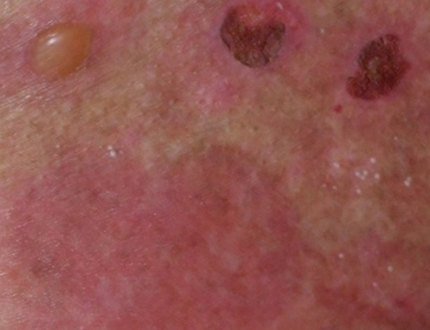
Bullous pemphigoid (BP) is the most prevalent autoimmune disorder characterized by subepidermal blistering, primarily affecting older adults. While BP primarily presents with tense bullae on the extremities and trunk, milia formation during recovery is a rare but notable occurrence. We report an 86-year-old Thai male diagnosed with BP who developed milia on the trunk and extremities during the recovery phase. Diagnosis was confirmed by clinical findings, histopathology, and immunological studies, which showed subepidermal blistering, elevated anti-BP180 levels, and epidermal-side IgG deposition. Treatment included prednisolone, doxycycline, nicotinamide, and topical clobetasol, with lesion improvement observed.
เอกสารอ้างอิง
Furumura M, Hashimoto T. Bullous pemphigoid with prominent milium formation. Acta Dermatovenerol Croat 2013;21:35-8.
Vernal S, de Oliveira EV, Filho RB, et al. Bullous pemphigoid and milia: prevalence and clinical laboratory findings in a Brazilian sample. An Bras Dermatol 2022;97:435-42.
Amin S, Fiore CT, Paek SY. Milia within resolving bullous pemphigoid lesions. Proc (Bayl Univ Med Cent) 2019;32:90-92.
Uchida S, Oiso N, Koga H, et al. Refractory bullous pemphigoid leaving numerous milia during recovery. J Dermatol 2014;41:1003-5.
Patsatsi A, Uy CDC, Murrell DF. Multiple milia formation in blistering diseases. Int J Womens Dermatol 2020;6:199-202.
Zhang LW, Li L, Chen T, Wang WJ, Fu LX, He L. Appearance of prominent milia as secondary lesions during recovery of refractory bullous pemphigoid: a case report. Int J Dermatol Venereol 2018;1:253-5.
Meryem Elomari Alaoui, Amani Fliti, Nadia Ismaili, Mariame Meziane, Laila Benzekri, Karima Senouci. Eruptive milia in a patient with bullous pemphigoid. J Case Rep Sci Images 2024;6:14-15.
Baican C, Candrea E, Baican A, Vasilovici A, Vornicescu D, Danescu S. Localized bullous pemphigoid associated with multiple milia formation - an atypical presentation. HVM Bioflux 2019;11:87-90.
Ding S, Deng Q, Xiang Y, Chen J, Huang J, Lu J. Bullous pemphigoid associated with milia, increased serum IgE, autoantibodies against desmogleins, and refractory treatment in a young patient. An Bras Dermatol 2017;92:34-6.
Kumudhini S, Rao R, Pai K, Shetty S, Pai S. Extensive milia formation in a young woman with bullous pemphigoid. Indian J Dermatol Venereol Leprol 2018;84:248.
Banfield CC, Wojnarowska F, Allen J, George S, Venning VA, Welsh KI. The association of HLA-DQ7 with bullous pemphigoid is restricted to men. Br J Dermatol 1998;138:1085-90.
Prost C, Fressinaud C, Ingen-Housz-Oro S. Milia en plaque after bullous pemphigoid: An unusual sequela. Cureus 2021;13:e314067.
ดาวน์โหลด
เผยแพร่แล้ว
รูปแบบการอ้างอิง
ฉบับ
ประเภทบทความ
สัญญาอนุญาต
ลิขสิทธิ์ (c) 2026 วารสารโรคผิวหนัง

อนุญาตภายใต้เงื่อนไข Creative Commons Attribution-NonCommercial-NoDerivatives 4.0 International License.
เนื้อหาและข้อมูลในบทความที่ลงตีพิมพ์ในวารสารโรคผิวหนัง ถือเป็นข้อคิดเห็นและความรับผิดชอบของผู้เขียนบทความโดยตรงซึ่งกองบรรณาธิการวารสาร ไม่จำเป็นต้องเห็นด้วย หรือร่วมรับผิดชอบใดๆ
บทความ ข้อมูล เนื้อหา รูปภาพ ฯลฯ ที่ได้รับการตีพิมพ์ในวารสารโรคผิวหนัง ถือเป็นลิขสิทธิ์ของวารสารฯ หากบุคคลหรือหน่วยงานใดต้องการนำทั้งหมดหรือส่วนหนึ่งส่วนใดไปเผยแพร่ต่อหรือเพื่อกระทำการใดๆ จะต้องได้รับอนุญาตเป็นลายลักอักษรจากบรรณาธิการวารสารโรคผิวหนังก่อนเท่านั้น

