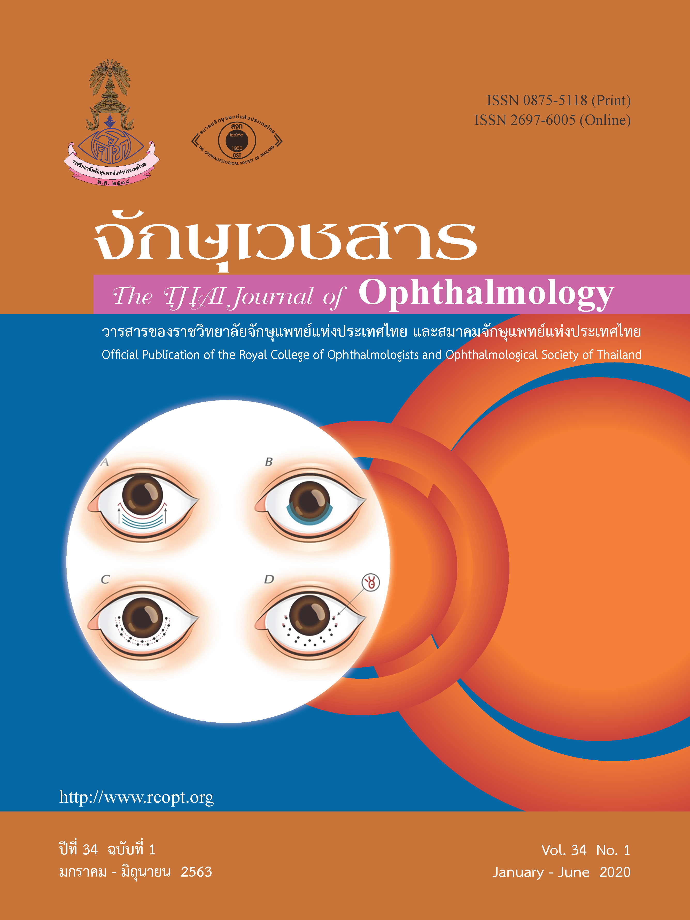
Conjunctivochalasis
Keywords:
ConjunctivochalasisAbstract
Conjunctivochalasis is an ocular condition characterized by loose, redundant, nonedematous conjunctiva. Although the prevalence of conjunctivochalsis in Thailand remains unknown, it has been documented that conjunctivochalsis increases in incidence and severity with age. This condition has also been found more frequently in patients with thyroid disease, dry eye, anterior blepharitis, meibomian gland dysfunction (MGD), and contact lens wear. Conjunctivochalasis usually occurs bilaterally, but it is often overlooked because the majority of cases are asymptomatic. As the disease progresses, a patient can become symptomatic. Common symptoms include ocular irritation, foreign body sensation, discomfort, dryness, and tearing. These symptoms are non-specific and similar to those of other more common ocular surface diseases such as dry eye disease which could be found concomitantly. The diagnosis of conjunctivochalsis is simply made by increasing awareness of its existence and thorough slit-lamp examination of the ocular surface, particularly in patients presented with chronic ocular surface disorders and poorly responded to medications. No treatment is necessary if the patient is asymptomatic. Once the patient develops significant symptoms, medical therapy is initiated with ocular lubricants, topical antihistamines, or topical steroids. Other components of concomitant ocular surface diseases, including allergic conjunctivitis, dry eye disease, and MGD should be concurrently managed. In cases where medical treatments fail to control the patient’s symptoms, surgical intervention is indicated. Conjunctivochalasis surgery is safe and effective and has an excellent prognosis. The surgery can help reduce or eliminate the symptoms over the long-term.
References
Di Pascuale MA, Espana EM, Kawakita T, et al. Clinical characteristics of conjunctivochalasis with or without aqueous tear deficiency. Br J Ophthalmol. 2004;88:388-92.
Mimura T, Yamagami S, Usui T, et al. Changes of conjunctivochalasis with age in a hospital-based study. Am J Ophthalmol. 2009;147:171-7.
Zhang X, Li Q, Zou H, et al. Assessing the severity of conjunctivochalasis in a senile population: a community-based epidemiology study in Shanghai, China. BMC Public Health. 2011;11:198. doi: 10.1186/ 1471-2458-11-198.
Mimura T, Usui T, Yamamoto H, et al. Conjunctivochalasis and contact lenses. Am J Ophthalmol. 2009;148:20-5.
de Almeida SF, de Sousa LB, Vieira LA, et al. Clinic-cytologic study of conjunctivochalasis and its relation to thyroid autoimmune diseases: prospective cohort study. Cornea. 2006;25:789-93.
Höh H, Schirra F, Kienecker C, et al. Lid-parallel conjunctival folds are a sure diagnostic sign of dry eye. Ophthalmologe. 1995;92:802-8.
Meller D, Tseng SC. Conjunctivochalasis: literature review and possible pathophysiology. Survey of Ophthalmology. 1998;43:225-32.
Li DQ, Meller D, Liu Y, et al. Overexpression of MMP-1 and MMP-3 by cultured conjunctivochalasis fibroblasts. Invest Ophthalmol Vis Sci. 2000;41:404-10.
Watanabe A, Yokoi N, Kinoshita S, et al. Clinicopathologic study of conjunctivochalasis. Cornea 2004;23:294-8.
Yokoi N, Komuro A, Nishii M, et al. Clinical impact of conjunctivochalasis on the ocular surface. Cornea 2005;24(8 Suppl):S24-S31.
Hara S, Kojima T, Ishida R, et al. Evaluation of tear stability after surgery for conjunctivochalasis. Optom Vis Sci. 2011;88:1112-8.
Wang S, Ke M, Cai X, et al. An improved surgical method to correct conjunctivochalasis: conjunctival semiperitomy based on corneal limbus with subconjunctival cauterization. Can J Ophthalmol. 2012;47:418-22.
Doss LR, Doss EL, Doss RP. Paste-pinch-cut conjunctivoplasty: subconjunctival fibrin sealant injection in the repair of conjunctivochalasis. Cornea 2012;31:959-62.
Georgiadis NS, Terzidou CD. Epiphora caused by conjunctivochalasis: treatment with transplantation of preserved human amniotic membrane. Cornea 2001;20:619-21.
Kheirkhah A, Casas V, Blanco G, et al. Amniotic membrane transplantation with fibrin glue for conjunctivochalasis. Am J Ophthalmol 2007;144:311-3.
Otaka I, Kyu N. A new surgical technique for management of conjunctivochalasis. Am J Ophthalmol 2000;129:385-7.
Downloads
Published
Issue
Section
License
The Thai Journal of Ophthalmology (TJO) is a peer-reviewed, scientific journal published biannually for the Royal College of Ophthalmologists of Thailand. The objectives of the journal is to provide up to date scientific knowledge in the field of ophthalmology, provide ophthalmologists with continuing education, promote cooperation, and sharing of opinion among readers.
The copyright of the published article belongs to the Thai Journal of Ophthalmology. However the content, ideas and the opinions in the article are from the author(s). The editorial board does not have to agree with the authors’ ideas and opinions.
The authors or readers may contact the editorial board via email at admin@rcopt.org.


