Distinguishing Lipid-Poor Adrenal Adenoma from Non-Adenomatous Lesions: Accuracy of Identification and the Most Appropriate Contrast Washout Index Using the Contrast-Enhanced Washout Multidetector CT 10-Minute Delayed Imaging Protocol
Keywords:
10-minute contrast washout CT, Adrenal adenoma, Absolute percentage washout, Lipid poor adenoma, Relative percentage washoutAbstract
Background: Because of the contrast washout characteristic of adrenal adenoma, Siriraj Hospital uses the 10-minute delayed imaging computed tomography (CT) protocol to evaluate adrenal masses. No reports in Thailand have described the performance of this protocol to date.
Objective: This study was performed to identify the accuracy of identification and the most appropriate contrast washout index to distinguish lipid-poor adrenal adenoma from non-adenomatous lesions using the 10-minute delayed imaging CT protocol.
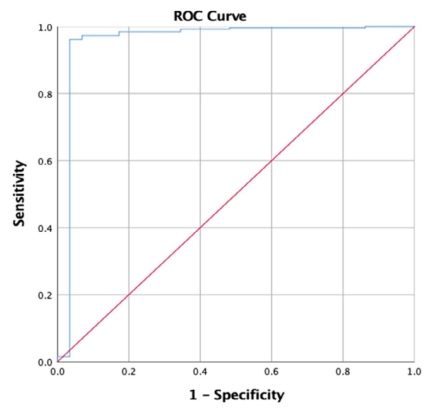
Materials and Methods: This retrospective review involved patients who underwent the CT adrenal protocol with 10-minute delayed imaging at Siriraj Hospital from January 2005 to December 2017. In total, 285 adrenal masses that were smaller than 4 cm and had a poor lipid component (density of >10 HU) were assessed in 261 patients who were given a pathologic diagnosis or underwent follow-up imaging. Non-contrast images were obtained before intravenous contrast administration with an 80-second and 10-minute scan delay. The absolute percentage washout (APW) and relative percentage washout (RPW) of the adrenal masses were calculated. Receiver operating characteristic analysis was performed to evaluate the protocol performance and the most appropriate contrast washout value with which to identify lipid-poor adrenal adenoma.
Results: The test appeared to be the most accurate when using an APW of 47% and RPW of 34%. The APW of 47% showed a sensitivity of 96.5%, specificity of 89.3%, and accuracy of 95.8% (p < 0.001), while the RPW of 34% showed a sensitivity of 89.5%, specificity of 96.4%, and accuracy of 90.2% (p < 0.001).
Conclusion: An APW of 47% and RPW of 34% were the most appropriate washout indexes, offering high accuracy to distinguish lipid-poor adrenal adenoma from non-adenomatous lesions.
Downloads
References
Song JH, Chaudhry FS, Mayo-Smith WW. The incidental adrenal mass on CT: prevalence of adrenal disease in 1,049 consecutive adrenal masses in patients with no known malignancy. AJR Am J Roentgenol. 2008; 190(5):1163-1168. doi:10.2214/AJR.07.2799 https://doi.org/10.2214/AJR.07.2799 PMid:18430826
Blake MA, Kalra MK, Sweeney AT, et al. Distinguishing benign from malignant adrenal masses: multi-detector row CT protocol with 10-minute delay. Radiology. 2006;238(2): 578-585. doi:10.1148/radiol.2382041514 https://doi.org/10.1148/radiol.2382041514 PMid:16371582
Patel MN, Rajpura HKK, Solanki RN, Patel H. Distinguishing benign lesion from malignant adrenal masses by CT scan with 15 minutes delayed protocol. J Evid Based Med Healthc. 2018;5(38):2747-2751. doi:10.18410/jebmh/ 2018/563 https://doi.org/10.18410/jebmh/2018/563
Johnson PT, Horton KM, Fishman EK. Adrenal Imaging with multidetector CT: evidence-based protocol optimization and interpretative practice.Radiographics. 2009;29(5):1319-1331. doi:10.1148/rg.295095026 https://doi.org/10.1148/rg.295095026 PMid:19755598
Peña CS, Boland GW, Hahn PF, Lee MJ, Mueller PR. Characterization of indeterminate (lipid-poor) adrenal masses: use of washout characteristics at contrast-enhanced CT. Radiology. 2000;217(3):798-802. doi:10.1148/ radiology.217.3.r00dc29798 https://doi.org/10.1148/radiology.217.3.r00dc29798 PMid:11110946
Szolar DH, Korobkin M, Reittner P, et al. Adrenocortical carcinomas and adrenal pheochromocytomas: mass and enhancement loss evaluation at delayed contrast-enhanced CT. Radiology. 2005;234(2):479-485. doi:10.1148/ radiol.2342031876 https://doi.org/10.1148/radiol.2342031876 PMid:15671003
Sangwaiya MJ, Boland GW, Cronin CG, Blake MA, Halpern EF, Hahn PF. Incidental adrenal lesions: accuracy of characterization with contrast-enhanced washout multidetector CT--10-minute delayed imaging protocol revisited in a large patient cohort. Radiology. 2010;256(2):504-510. doi:10.1148/radiol. 10091386 https://doi.org/10.1148/radiol.10091386 PMid:20656838

Downloads
Published
How to Cite
Issue
Section
License
Copyright (c) 2022 Chulabhorn Royal Academy

This work is licensed under a Creative Commons Attribution-NonCommercial-NoDerivatives 4.0 International License.
Copyright and Disclaimer
Articles published in this journal are the copyright of Chulabhorn Royal Academy.
The opinions expressed in each article are those of the individual authors and do not necessarily reflect the views of Chulabhorn Royal Academy or any other faculty members of the Academy. The authors are fully responsible for all content in their respective articles. In the event of any errors or inaccuracies, the responsibility lies solely with the individual authors.


