
Radiation Dose Reduction in Whole Brain Perfusion Computed Tomography Using 320-detector Computed Tomography
Keywords:
320-detector CT, radiation dose, whole- brain perfusion CTAbstract
Background: Radiation dose reduction in whole-brain perfusion computed tomography (CT) can be performed using various methods. We used a protocol with approximately 30% tube current of the original control protocol (Protocol 1) and a reduced total scan volume protocol (Protocol 2).
Methods: We conducted a retrospective consecutively analysis of whole-brain perfusion CT in 15 adult patients using the control protocol. A further 10 patients underwent protocol 1, and another 10 patients underwent protocol 2. We performed quantitative analyses and assessed the diagnostic image quality of the parametric map images. The percentage of radiation dose reductions was calculated.
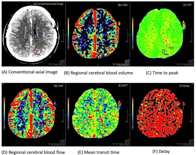
Results: The gray and white matter signal intensities were higher in the cerebral blood volume and flow maps of protocol 1 than protocol 2, whereas this opposite results pattern was reversed for the time to peak and mean transit time maps. The signal-to-noise ratios of the gray and white matter for protocol 1 was inferior to that of protocol 2 for all perfusion parameters. No significant quantitative differences in parametric maps were found between the control protocol and protocol 1 or 2. The differences in the radiation dose reduction between protocols 1 and 2 and the control protocol were 33.23% and 19.95%, respectively. The effective dose of protocol 1 was reduced to approximately half that of the control protocol.
Conclusions: Radiation dose was significantly lower in protocol 1 than in the control protocol while providing comparable parametric image quality in regard to both the gray and white matter signal and signal-to-noise ratios of the parametric maps.
Downloads
References
Sahani DV. Perfusion CT: An Overview Of Technique And Clinical Applications. 2010.
Corcuera-Solano I, McLellan AM, Doshi AH, Pawha PS, Tanenbaum LN. Whole-brain adaptive 70-kVp perfusion imaging with variable and extended sampling improves quality and consistency while reducing dose. AJNR Am J Neuroradiol. 2014;35(11):2045-2051. https://doi.org/10.3174/ajnr.A4043 PMid:25034777 PMCid:PMC7965192
Shankar JJ, Lum C. Whole brain CT perfusion on a 320-slice CT scanner. Indian J Radiol Imaging. 2011;21(3):209-214. https://doi.org/10.4103/0971-3026.85370 PMid:22013297 PMCid:PMC3190494
Wintermark M, Lev MH. FDA investigates the safety of brain perfusion CT. AJNR Am J Neuroradiol. 2010;31(1):2-3. https://doi.org/10.3174/ajnr
Konstas AA, Goldmakher GV, Lee TY, Lev MH. Theoretic basis and technical implementations of CT perfusion in acute ischemic stroke, part 1: Theoretic basis. AJNR Am J Neuroradiol. 2009;30(4):662-668. https://doi.org/10.3174/ajnr.A1487 PMid:19270105 PMCid:PMC7051780
Fieselmann A, Kowarschik M, Ganguly A, Hornegger J, Fahrig R. Deconvolution-Based CT and MR Brain Perfusion Measurement: Theoretical Model Revisited and Practical Implementation Details. Int J Biomed Imaging. 2011;2011:467563. https://doi.org/10.1155/2011/467563 PMid:21904538 PMCid:PMC3166726
Kämena A, Streitparth F, Grieser C, et al. Dynamic perfusion CT: optimizing the temporal resolution for the calculation of perfusion CT parameters in stroke patients. Eur J Radiol. 2007;64(1):111-118. https://doi.org/10.1016/j.ejrad.2007.02.025 PMid:17383135
Wintermark M, Maeder P, Verdun FR, et al. Using 80 kVp versus 120 kVp in perfusion CT measurement of regional cerebral blood flow. AJNR Am J Neuroradiol. 2000; 21(10):1881-1884.
Hirata M, Sugawara Y, Fukutomi Y, et al. Measurement of radiation dose in cerebral CT perfusion study. Radiat Med. 2005; 23(2):97-103.
Othman AE, Brockmann C, Yang Z, et al. Effects of radiation dose reduction in Volume Perfusion CT imaging of acute ischemic stroke. Eur Radiol. 2015;25(12):3415-3422. https://doi.org/10.1007/s00330-015-3763-7 PMid:25903716
Angel E. SVD + Dynamic Volume CT : Delay Insensitive Brain Perfusion Analysis. 2010.
Niesten JM, van der Schaaf IC, Riordan AJ, et al. Radiation dose reduction in cerebral CT perfusion imaging using iterative reconstruction. Eur Radiol. 2014;24(2):484-493. https://doi.org/10.1007/s00330-013-3042-4 PMid:24126642
Shrimpton PC, Hillier MC, Lewis MA, Dunn M. National survey of doses from CT in the UK: 2003. Br J Radiol. 2006;79(948): 968-980. https://doi.org/10.1259/bjr/93277434 PMid:17213302
Agency IAE. Dosimetry in Diagnostic Radiology for PaediatricPatients. 2013.
Cohnen M, Fischer H, Hamacher J, Lins E, Kötter R, Mödder U. CT of the head by use of reduced current and kilovoltage: relationship between image quality and dose reduction. AJNR Am J Neuroradiol. 2000;21(9):1654-1660.
Wintermark M, Smith WS, Ko NU, Quist M, Schnyder P, Dillon WP. Dynamic perfusion CT: optimizing the temporal resolution and contrast volume for calculation of perfusion CT parameters in stroke patients. AJNR Am J Neuroradiol. 2004;25(5):720-729.
Abels B, Klotz E, Tomandl BF, Villablanca JP, Kloska SP, Lell MM. CT perfusion in acute ischemic stroke: a comparison of 2-second and 1-second temporal resolution. AJNR Am J Neuroradiol. 2011;32(9):1632-1639. https://doi.org/10.3174/ajnr.A2576 PMid:21816919 PMCid:PMC7965399
Admontree S, Krisanachinda A, Laothamatas J, Trinavarat P. Radiation dose on whole brain computed tomography in comprehensive stroke imaging using axial volumetric 320- detector CT, Chulalongkorn University; 2013.
Siebert E, Bohner G, Dewey M, et al. 320-slice CT neuroimaging: initial clinical experience and image quality evaluation. Br J Radiol. 2009;82(979):561-570. https://doi.org/10.1259/bjr/27721218 PMid:19221186
Diekmann S, Siebert E, Juran R, et al. Dose exposure of patients undergoing comprehensive stroke imaging by multidetector-row CT: comparison of 320-detector row and 64-detector row CT scanners. AJNR Am J Neuroradiol. 2010;31(6):1003-1009. https://doi.org/10.3174/ajnr.A1971 PMid:20110373 PMCid:PMC7963949
Downloads
Published
How to Cite
Issue
Section
License
Copyright (c) 2022 Chulabhorn Royal Academy

This work is licensed under a Creative Commons Attribution-NonCommercial-NoDerivatives 4.0 International License.
Copyright and Disclaimer
Articles published in this journal are the copyright of Chulabhorn Royal Academy.
The opinions expressed in each article are those of the individual authors and do not necessarily reflect the views of Chulabhorn Royal Academy or any other faculty members of the Academy. The authors are fully responsible for all content in their respective articles. In the event of any errors or inaccuracies, the responsibility lies solely with the individual authors.

