
Determination radiation exposure rate at various distances and time for myocardial blood flow study using oxygen-15 water PET/CT imaging
Keywords:
15O-water, PET imaging, myocardial blood flow, radiation exposure rateAbstract
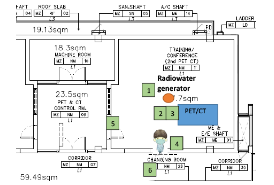
Background: Oxygen-15-labelled water is considered a gold standard for myocardial blood flow quantification. The present study aimed to measure radiation exposure rates from patients study with 15O-water myocardial blood flow (MBF) positron emission tomography/computed tomography (PET/CT) scans, and to assess occupational and public exposure. Methods: Radiation exposure rates from 15O-water were measured during both rest and stress study. The measurements were obtained at 2.15 min after 15O-water synthesis 1m away from the generator and 5 cm from the xiphoid level of each patient at time-points of 3.0, 8.30, and 15.0 min. Radiation exposure in the adjacent PET/CT imaging rooms was also surveyed. Results: The mean administered activities were 552.27±30.87MBq and 548.59±35.77MBq during rest and stress, respectively. During the 15O-water synthesis, the mean radiation exposure rates were 242.90±63.68µSv/h at rest and 254.60±56.92µSv/h during stress. The mean radiation exposure rates at 3.0, 8.30, and 15.0 min were 1305.90±419.64 µSv/h, 121.82±22.89 µSv/h, and 14.62±3.44µSv/h at rest, and 1320.07±451.83µSv/h, 124.49±24.48µSv/h, and 12.50±3.76µSv/h during stress, respectively. Radiation exposure rates in the PET/CT control room and at all locations outside the imaging room were within the background range. Conclusion: During15O-water PET/CT examinations of MBF patients. The radiation exposure rates from the patients provided the radiation safety for both radiation worker and the public according to the guidelines provided by the International Commission on Radiological Protection and the Office of Atoms for Peace. However the staff should be spend less time near the radiowater generator (this includes the patients after injected radiotracer and imaging), and increasing distance from the radioactive source and patients during diagnosis for deceasing the radiation exposure.
Downloads
References
Hsu B. PET tracers and techniques for measuring myocardial blood flow in patients with coronary artery disease. J Biomed Res. 2013;27(6):452-459. doi:10.7555/JBR.27.20130136
Manabe O, Kikuchi T, Scholte AJHA, et al. Radiopharmaceutical tracers for cardiac imaging [published correction appears in J Nucl Cardiol. 2018 Jan 24;:]. J Nucl Cardiol. 2018;25(4):1204-1236. doi:10.1007/s12350-017-1131-5
Smith H. The 1990 Recommendations of the International Commission on Radiological Protection. ICRP Publication 6. New York. 1991. Volume 21. No.1-3.
The 2007 Recommendations of the International Commission on Radiological Protection, ICRP Publication 103. 2007. http://www.nrc.gov/ docs/ML1233/ML12338A682.pdf. Accessed 30 Dec 2021.
International Atomic Energy Agency. Cyclotron Produced Radionuclides: Physical Characteristics and Production Methods. TRS no. 468. Vienna. 2009.https://wwwpub.iaea.org/mtcd/publications/pdf/trs468_web.pdf. Accessed 30 Dec 2021.
Anderson JA and Mathews D. Site planning and Radiation safety in PET facility. 2021. http://www.aapm.org/meetings/02AM/pdf/8418-39272.pdf. Accessed 30 Dec 2021.
Manabe O, Naya M, Aikawa T, Yoshinaga K. 15O-labeled Water is the Best Myocardial Blood Flow Tracer for Precise MBF Quantification. Annals of Nuclear Cardiology. 2019;5(1):69-72.
Nakazato R, Berman DS, Alexanderson E, Slomka P. Myocardial perfusion imaging with PET. Imaging Med. 2013;5(1):35-46. doi:10.2217/iim.13.1
Ribeiro ASF, Husson O, Drey N, et al. Radiation exposure awareness from patients undergoing nuclear medicine diagnostic 99mTc-MDP bone scans and 2-deoxy-2-(18F) fluoro-D-glucose PET/computed tomography scans. Nucl Med Commun. 2020;41(6):582-588. doi:10.1097/MNM.0000000 000001177
Mettler FA Jr, Mahesh M, Bhargavan-Chatfield M, et al. Patient Exposure from Radiologic and Nuclear Medicine Procedures in the United States: Procedure Volume and Effective Dose for the Period 2006-2016. Radiology. 2020;295(2):418-427. doi:10.1148/radiol.2020192256
Berberoglu K. External radiation exposure rate after 18F-FDG PET/CT examination. Radioprotection. 2019;54(2):113-6.
Office of Atoms for Peace. Ministry Regulations for Radiation Safety. 2561. http://www.ratchakitcha.soc.go.th/DATA/PDF/2561/A/079/T_0009.PDF. Accessed 30 Dec 2021.
Mountford PJ, O’Doherty MJ. Exposure of critical groups to nuclear medicine patients. Appl Radiat Isot. 1999;50(1):89-111.
Downloads
Published
How to Cite
Issue
Section
License
Copyright (c) 2023 Chulabhorn Royal Academy

This work is licensed under a Creative Commons Attribution-NonCommercial-NoDerivatives 4.0 International License.
Copyright and Disclaimer
Articles published in this journal are the copyright of Chulabhorn Royal Academy.
The opinions expressed in each article are those of the individual authors and do not necessarily reflect the views of Chulabhorn Royal Academy or any other faculty members of the Academy. The authors are fully responsible for all content in their respective articles. In the event of any errors or inaccuracies, the responsibility lies solely with the individual authors.

