
Tinea Capitis Incognito in Adult: A Case Report
Keywords:
tinea capitis, adult, tinea incognitAbstract
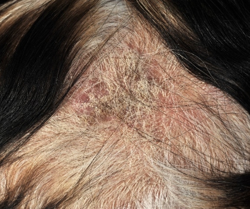
Tinea capitis is a superficial fungal infection of the hair and scalp caused by dermatophytes, usually Trichophyton and Microsporum species.1 Tinea capitis is common in children, while it occurs in adults who are immunocompromised or postmenopausal. Clinical manifestation of tinea capitis is highly variable, including grey patch, black dot, pustules, kerion, and favus. Tinea capitis in adult patients may have different clinical manifestations from those in children, which lead to the difficulty in diagnosis and consequently delay in treatment. Moreover, the lesions that have been treated with corticosteroids could potentially change to mimic other scalp diseases. We reported a 64-year-old woman with tinea capitis mimicking scalp dermatitis and receiving topical steroid. Trichoscopy and wood lamp’s examination help to make the diagnosis. KOH preparation and fungal culture are valuable to confirm the diagnosis and determine the causative organisms.
References
2. Khosravi AR, Shokri H, Vahedi G. Factors in etiology and predisposition of adult tinea capitis and review of published literature. Mycopathologia. 2016;181:371-8.
3. Ahmed SM, Rather SR, Kousar H, Bukhari S. Tinea capitis in adults: not so rare. Int J Res Med Sci 2016;4:5426-9.
4. Dutta B, Rasul ES, Boro B. Clinico-epidemiological study of tinea incognito with microbiological correlation. Indian J Dermatol Venereol Leprol.2017;83:326-31.
5. Ansar A, Farshchian M, Nazeri H, Ghiasian SA. Clinico-epidemiological and mycological aspects of tinea incognito in Iran: A 16-year study. Med Mycol J.2011;52:25-32.
6. Kastelan M, Prpic Massari L, Simonic E, Gruber F. Tinea incognito due to Microsporum canis in a 76- year-old woman. Wien Klin Wochenschr.2007;119:455.
7. Campos S, Brasileiro A, Galhardas C, Apetato M, Cabete J, Serrão V, et al. Follow-up of tinea capitis with trichoscopy: a prospective clinical study. J Eur Acad Dermatol Venereol. 2017;31:478-80.
8. Richarz NA, Barboza L, Monsonís M, González- Enseñat MA, Vicente A. Trichoscopy helps to predict the time point of clinical cure of tinea capitis. Australas J Dermatol.2018.
9. Gómez Moyano E, Crespo Erchiga V, Martínez Pilar L, Martinez García S. Correlation between dermoscopy and direct microscopy of morse code hairs in tinea incognito. J Am Acad Dermatol. 2016;74:7-8.
10. Chen X, Jiang X, Yang M, Bennett C, González U, Lin X, et al. Systemic antifungal therapy for tinea capitis in children: An abridged Cochrane Review. J Am Acad Dermatol.2017;76:368-74.
Downloads
Published
How to Cite
Issue
Section
License
เนื้อหาและข้อมูลในบทความที่ลงตีพิมพ์ในวารสารโรคผิวหนัง ถือเป็นข้อคิดเห็นและความรับผิดชอบของผู้เขียนบทความโดยตรงซึ่งกองบรรณาธิการวารสาร ไม่จำเป็นต้องเห็นด้วย หรือร่วมรับผิดชอบใดๆ
บทความ ข้อมูล เนื้อหา รูปภาพ ฯลฯ ที่ได้รับการตีพิมพ์ในวารสารโรคผิวหนัง ถือเป็นลิขสิทธิ์ของวารสารฯ หากบุคคลหรือหน่วยงานใดต้องการนำทั้งหมดหรือส่วนหนึ่งส่วนใดไปเผยแพร่ต่อหรือเพื่อกระทำการใดๆ จะต้องได้รับอนุญาตเป็นลายลักอักษรจากบรรณาธิการวารสารโรคผิวหนังก่อนเท่านั้น

