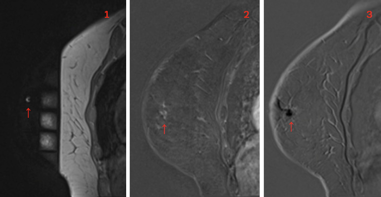
Magnetic Resonance Imaging-Guided Vacuum-Assisted Breast Biopsy in Chulabhorn hospital
Keywords:
Breast biopsy, complications, Vacuum assisted breast biopsyAbstract
MRI of the breast is currently used for high-risk screening, preoperative staging, and post treatment follow-up. Following the increasing clinical use of breast MRI, there has been a concomitant increase in the number of MRI-guided intervention for lesions only visible with breast MRI. Despite the highest sensitivity of MRI for breast cancer detection among current clinical imaging modalities, its a lower specificity has been noted. Besides, MRI-guided breast biopsy is often required for diagnosis and patient management. Thus, the purpose of this article is to review indications, contraindications, breast MRI biopsy equipment, the workflow and technique of breast MRI-guided biopsy in Chulabhorn hospital.
Downloads
References
Peters NH, Borel Rinkes IH, Zuithoff NP, Mali WP, Moons KG, Peeters PH. Metaanalysis of MR imaging in the diagnosis of breast lesions. Radiology. 2008;246(1): 116-124. doi:10.1148/radiol.2461061298
Schnall MD, Blume J, Bluemke DA, et al. Diagnostic architectural and dynamic features at breast MR imaging: multicenter study. Radiology. 2006;238(1):42-53. doi:10.1148/radiol.2381042117
LaTrenta LR, Menell JH, Morris EA, Abramson AF, Dershaw DD, Liberman L. Breast lesions detected with MR imaging: utility and histopathologic importance of identification with US. Radiology. 2003;227(3):856-861. doi:10.1148/radiol.2273012210
Meissnitzer M, Dershaw DD, Lee CH, Morris EA. Targeted ultrasound of the breast in women with abnormal MRI findings for whom biopsy has been recommended. AJR Am J Roentgenol. 2009;193(4):1025-1029.doi:10.2214/AJR.09.2480
Abe H, Schmidt RA, Shah RN, et al. MRdirected (“Second-Look”) ultrasound examination for breast lesions detected initially on MRI: MR and sonographic findings. AJR Am J Roentgenol. 2010; 194(2):370-377. doi:10.2214/AJR.09.2707
Beran L, Liang W, Nims T, Paquelet J, Sickle-Santanello B. Correlation of targeted ultrasound with magnetic resonance imaging abnormalities of the breast. Am J Surg. 2005;190(4):592-594. doi:10.1016/j.amjsurg.2005.06.019
Heywang-Köbrunner SH, Sinnatamby R, Lebeau A, et al. Interdisciplinary consensus on the uses and technique of MR-guided vacuum-assisted breast biopsy (VAB): results of a European consensus meeting. Eur J Radiol. 2009;72(2):289-294. doi:10.1016/j.ejrad.2008.07.010
Eby PR, Lehman C. MRI-guided breast interventions. Semin Ultrasound CT MR. 2006;27(4):339-350. doi:10.1053/j.sult.2006.05.008
Liberman L, Bracero N, Morris E, Thornton C, Dershaw DD. MRI-guided 9-gauge vacuum-assisted breast biopsy: initial clinical experience. AJR Am J Roentgenol. 2005;185(1):183-193. doi:10.2214/ajr.185.1.01850183
Mahoney MC. Initial clinical experience with a new MRI vacuum-assisted breast biopsy device. J Magn Reson Imaging. 2008;28(4):900-905. doi:10.1002/jmri.21549
Perlet C, Heywang-Kobrunner SH, Heinig A, et al. Magnetic resonanceguided, vacuum-assisted breast biopsy: results from a European multicenter study of 538 lesions. Cancer. 2006;106(5): 982-990. doi:10.1002/cncr.21720
Hefler L, Casselman J, Amaya B, et al. Follow-up of breast lesions detected by MRI not biopsied due to absent enhancement of contrast medium. Eur Radiol. 2003;13 (2):344-346. doi:10.1007/s00330-002-1713-7
Liberman L, Morris EA, Dershaw DD, Thornton CM, Van Zee KJ, Tan LK. Fast MRI-guided vacuum-assisted breast biopsy: initial experience. AJR Am J Roentgenol. 2003;181(5):1283-1293. doi: 10.2214/ajr.181.5.1811283
D’Orsi CJ, Sickles EA, Mendelson EB, Morris EA. ACR BI-RADS Atlas, breast imaging reporting and data system. fifth ed: American College of Radiology; 2013.
Gilbert FJ, Warren RM, Kwan-Lim G, et al. Cancers in BRCA1 and BRCA2 carriers and in women at high risk for breast cancer: MR imaging and mammographic features. Radiology. 2009;252(2):358-368. doi:10.1148/radiol.2522081032
Sardanelli F, Podo F, D’Agnolo G, et al. Multicenter comparative multimodality surveillance of women at genetic-familial high risk for breast cancer (HIBCRIT study) : interim results. Radiology. 2007;242(3): 698-715. doi:10.1148/radiol.2423051965
Friedlander LC, Roth SO, Gavenonis SC. Results of MR imaging screening for breast cancer in high-risk patients with lobular carcinoma in situ. Radiology. 2011;261(2):421-427. doi:10.1148/radiol. 11103516
Somerville P, Seifert PJ, Destounis SV, Murphy PF, Young W. Anticoagulation and bleeding risk after core needle biopsy. AJR Am J Roentgenol. 2008;191(4) :1194-1197. doi:10.2214/AJR.07.3537
Han BK, Schnall MD, Orel SG, Rosen M. Outcome of MRI-guided breast biopsy. AJR Am J Roentgenol. 2008;191(6):1798-1804. doi:10.2214/AJR.07.2827
El Khouli RH, Macura KJ, Kamel IR, Bluemke DA, Jacobs MA. The effects of applying breast compression in dynamic contrast material-enhanced MR imaging. Radiology. 2014;272(1):79-90. doi:10. 1148/radiol.14131384
Brennan SB, Sung JS, Dershaw DD, Liberman L, Morris EA. Cancellation of MR imaging-guided breast biopsy due to lesion nonvisualization: frequency and follow-up. Radiology. 2011;261(1): 92-99. doi:10.1148/radiol.11100720
Johnson KS, Baker JA, Lee SS, Soo MS. Cancelation of MRI guided breast biopsies for suspicious breast lesions identified at 3.0 T MRI: reasons, rates, and outcomes. Acad Radiol. 2013;20(5) :569-575. doi:10.1016/j.acra.2013.01.005
Niell BL, Lee JM, Johansen C, Halpern EF, Rafferty EA. Patient outcomes in canceled MRI-guided breast biopsies. AJR Am J Roentgenol. 2014;202(1):223- doi:10.2214/AJR.12.10228.228

Downloads
Published
How to Cite
Issue
Section
License
Copyright (c) 2023 Chulabhorn Royal Academy

This work is licensed under a Creative Commons Attribution-NonCommercial-NoDerivatives 4.0 International License.
Copyright and Disclaimer
Articles published in this journal are the copyright of Chulabhorn Royal Academy.
The opinions expressed in each article are those of the individual authors and do not necessarily reflect the views of Chulabhorn Royal Academy or any other faculty members of the Academy. The authors are fully responsible for all content in their respective articles. In the event of any errors or inaccuracies, the responsibility lies solely with the individual authors.


