
การเจาะชิ้นเนื้อเต้านมด้วยเครื่องดูดระบบสุญญากาศ โดยใช้การตรวจคลื่นแม่เหล็กไฟฟ้าเป็นภาพนำตำแหน่งในโรงพยาบาลจุฬาภรณ์
คำสำคัญ:
การเจาะชิ้นเนื้อเต้านม, การใช้เครื่องเอ็มอาร์ไอเป็นภาพนำตำแหน่ง, เครื่องดูดชิ้นเนื้อเต้านมด้วยระบบสุญญากาศบทคัดย่อ
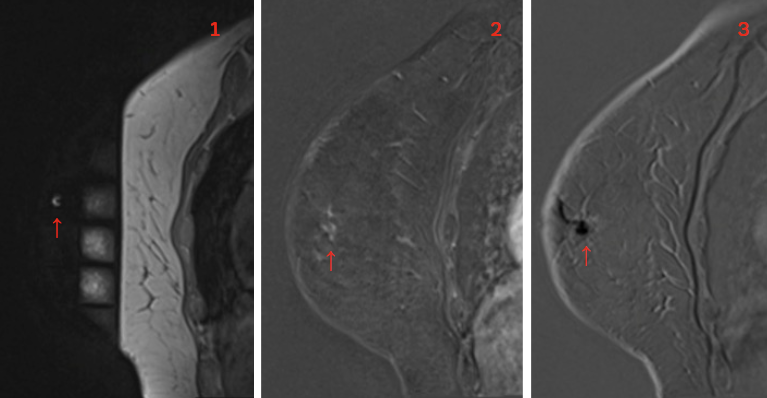
ปัจจุบันการตรวจคลื่นแม่เหล็กไฟฟ้า (เอ็มอาร์ไอ) เต้านมเป็นวิธีการตรวจที่ใช้กันอย่างแพร่หลาย เพื่อคัดกรองมะเร็งเต้านมในผู้หญิงที่มีความเสี่ยงสูง ใช้ในการตรวจผู้ป่วยมะเร็งเต้านมเพื่อวางแผนรักษา รวมถึง การตรวจติดตามผู้ป่วยมะเร็งเต้านมหลังให้การรักษา การตรวจเอ็มอาร์ไอเต้านมที่มากขึ้นนี้ ส่งผลให้มีการเจาะชิ้นเนื้อเต้านม โดยใช้เครื่องเอ็มอาร์ไอเป็นภาพนำตำแหน่งในกลุ่มรอยโรคที่ตรวจพบเฉพาะจากเครื่องเอ็มอาร์ไอเท่านั้นมากขึ้นไปด้วย ถึงแม้ว่าเอ็มอาร์ไอเต้านมจะมีความไวในการตรวจพบมะเร็งเต้านมมากกว่าวิธีการตรวจอื่น ๆ แต่ความจำาเพาะค่อนข้างตื่น ดังนั้น การเจาะชิ้นเนื้อเต้านมโดยใช้เครื่องเอ็มอาร์ไอเป็นภาพ นำตำแหน่งจึงมีความจำาเป็นเพื่อให้ผู้ป่วยได้รับวินิจฉัยโรคที่ถูกต้อง และเพื่อกำหนดแนวทางรักษาหรือการตรวจติดตามต่อไป วัตถุประสงค์ของบทความนี้ เพื่อทบทวนข้อบ่งชี้ ข้อห้าม รวมถึงอุปกรณ์ที่ใช้ ตลอดจน ขั้นตอนและเทคนิคในการเจาะชิ้นเนื้อเต้านมโดยใช้เครื่องเอ็มอาร์ไอเป็นภาพนำตำแหน่งในโรงพยาบาลจุฬาภรณ์
Downloads
เอกสารอ้างอิง
Peters NH, Borel Rinkes IH, Zuithoff NP, Mali WP, Moons KG, Peeters PH. Metaanalysis of MR imaging in the diagnosis of breast lesions. Radiology. 2008;246(1): 116-124. doi:10.1148/radiol.2461061298
Schnall MD, Blume J, Bluemke DA, et al. Diagnostic architectural and dynamic features at breast MR imaging: multicenter study. Radiology. 2006;238(1):42-53. doi:10.1148/radiol.2381042117
LaTrenta LR, Menell JH, Morris EA, Abramson AF, Dershaw DD, Liberman L. Breast lesions detected with MR imaging: utility and histopathologic importance of identification with US. Radiology. 2003;227(3):856-861. doi:10.1148/radiol.2273012210
Meissnitzer M, Dershaw DD, Lee CH, Morris EA. Targeted ultrasound of the breast in women with abnormal MRI findings for whom biopsy has been recommended. AJR Am J Roentgenol. 2009;193(4):1025-1029.doi:10.2214/AJR.09.2480
Abe H, Schmidt RA, Shah RN, et al. MRdirected (“Second-Look”) ultrasound examination for breast lesions detected initially on MRI: MR and sonographic findings. AJR Am J Roentgenol. 2010; 194(2):370-377. doi:10.2214/AJR.09.2707
Beran L, Liang W, Nims T, Paquelet J, Sickle-Santanello B. Correlation of targeted ultrasound with magnetic resonance imaging abnormalities of the breast. Am J Surg. 2005;190(4):592-594. doi:10.1016/j.amjsurg.2005.06.019
Heywang-Köbrunner SH, Sinnatamby R, Lebeau A, et al. Interdisciplinary consensus on the uses and technique of MR-guided vacuum-assisted breast biopsy (VAB): results of a European consensus meeting. Eur J Radiol. 2009;72(2):289-294. doi:10.1016/j.ejrad.2008.07.010
Eby PR, Lehman C. MRI-guided breast interventions. Semin Ultrasound CT MR. 2006;27(4):339-350. doi:10.1053/j.sult.2006.05.008
Liberman L, Bracero N, Morris E, Thornton C, Dershaw DD. MRI-guided 9-gauge vacuum-assisted breast biopsy: initial clinical experience. AJR Am J Roentgenol. 2005;185(1):183-193. doi:10.2214/ajr.185.1.01850183
Mahoney MC. Initial clinical experience with a new MRI vacuum-assisted breast biopsy device. J Magn Reson Imaging. 2008;28(4):900-905. doi:10.1002/jmri.21549
Perlet C, Heywang-Kobrunner SH, Heinig A, et al. Magnetic resonanceguided, vacuum-assisted breast biopsy: results from a European multicenter study of 538 lesions. Cancer. 2006;106(5): 982-990. doi:10.1002/cncr.21720
Hefler L, Casselman J, Amaya B, et al. Follow-up of breast lesions detected by MRI not biopsied due to absent enhancement of contrast medium. Eur Radiol. 2003;13 (2):344-346. doi:10.1007/s00330-002-1713-7
Liberman L, Morris EA, Dershaw DD, Thornton CM, Van Zee KJ, Tan LK. Fast MRI-guided vacuum-assisted breast biopsy: initial experience. AJR Am J Roentgenol. 2003;181(5):1283-1293. doi: 10.2214/ajr.181.5.1811283
D’Orsi CJ, Sickles EA, Mendelson EB, Morris EA. ACR BI-RADS Atlas, breast imaging reporting and data system. fifth ed: American College of Radiology; 2013.
Gilbert FJ, Warren RM, Kwan-Lim G, et al. Cancers in BRCA1 and BRCA2 carriers and in women at high risk for breast cancer: MR imaging and mammographic features. Radiology. 2009;252(2):358-368. doi:10.1148/radiol.2522081032
Sardanelli F, Podo F, D’Agnolo G, et al. Multicenter comparative multimodality surveillance of women at genetic-familial high risk for breast cancer (HIBCRIT study) : interim results. Radiology. 2007;242(3): 698-715. doi:10.1148/radiol.2423051965
Friedlander LC, Roth SO, Gavenonis SC. Results of MR imaging screening for breast cancer in high-risk patients with lobular carcinoma in situ. Radiology. 2011;261(2):421-427. doi:10.1148/radiol. 11103516
Somerville P, Seifert PJ, Destounis SV, Murphy PF, Young W. Anticoagulation and bleeding risk after core needle biopsy. AJR Am J Roentgenol. 2008;191(4) :1194-1197. doi:10.2214/AJR.07.3537
Han BK, Schnall MD, Orel SG, Rosen M. Outcome of MRI-guided breast biopsy. AJR Am J Roentgenol. 2008;191(6):1798-1804. doi:10.2214/AJR.07.2827
El Khouli RH, Macura KJ, Kamel IR, Bluemke DA, Jacobs MA. The effects of applying breast compression in dynamic contrast material-enhanced MR imaging. Radiology. 2014;272(1):79-90. doi:10. 1148/radiol.14131384
Brennan SB, Sung JS, Dershaw DD, Liberman L, Morris EA. Cancellation of MR imaging-guided breast biopsy due to lesion nonvisualization: frequency and follow-up. Radiology. 2011;261(1): 92-99. doi:10.1148/radiol.11100720
Johnson KS, Baker JA, Lee SS, Soo MS. Cancelation of MRI guided breast biopsies for suspicious breast lesions identified at 3.0 T MRI: reasons, rates, and outcomes. Acad Radiol. 2013;20(5) :569-575. doi:10.1016/j.acra.2013.01.005
Niell BL, Lee JM, Johansen C, Halpern EF, Rafferty EA. Patient outcomes in canceled MRI-guided breast biopsies. AJR Am J Roentgenol. 2014;202(1):223- doi:10.2214/AJR.12.10228.228
ดาวน์โหลด
เผยแพร่แล้ว
รูปแบบการอ้างอิง
ฉบับ
ประเภทบทความ
สัญญาอนุญาต
ลิขสิทธิ์ (c) 2023 ราชวิทยาลัยจุฬาภรณ์

อนุญาตภายใต้เงื่อนไข Creative Commons Attribution-NonCommercial-NoDerivatives 4.0 International License.
บทความที่ได้รับการตีพิมพ์เป็นลิขสิทธิ์ของราชวิทยาลัยจุฬาภรณ์
ข้อความที่ปรากฏในบทความแต่ละเรื่องในวารสารวิชาการเล่มนี้เป็นความคิดเห็นส่วนตัวของผู้เขียนแต่ละท่านไม่เกี่ยวข้องกับราชวิทยาลัยจุฬาภรณ์ และคณาจารย์ท่านอื่น ในราชวิทยาลัยฯ แต่อย่างใด ความรับผิดชอบองค์ประกอบทั้งหมดของบทความแต่ละเรื่องเป็นของผู้เขียนแต่ละท่าน หากมีความผิดพลาดใด ๆ ผู้เขียนแต่ละท่านจะรับผิดชอบบทความของตนเองแต่ผู้เดียว

