Effect of fluoride-containing resin sealant on the subsurface enamel microhardness of artificial incipient caries lesions: An in vitro study
Main Article Content
Abstract
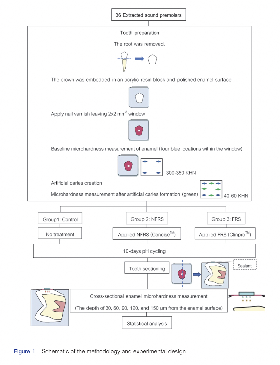
Objective: To evaluate the subsurface enamel microhardness at different depths on artificial incipient enamel caries using fluoride-releasing resin sealants.
Materials and Methods: Artificial enamel caries were created at the buccal surface of thirty-six extracted human premolars and randomly divided into three groups (n=12): Group 1: untreated group (control), Group 2: fluoride-releasing resin sealant (FRS, ClinproTM), and Group 3: non-fluoride releasing resin sealant (NFRS, ConciseTM). The sealant was placed on the buccal window (2x2x1 mm3) of the teeth in groups 2 and 3; and the groups underwent pH cycling for ten days. The teeth were sectioned, and the Knoop hardness number (KHN) was measured at 30-, 60-, 90-, 120-, and 150-µm deep from the enamel surface. The data were analyzed using one-way repeated ANOVA and pairwise comparisons with the Bonferroni test with a significance level of p < 0.05.
Results: At 30-µm depth, the enamel microhardness in the FRS group was the highest (159.62 ± 30.66 KHN) and statistically significantly higher than the NFRS (120.64 ± 38.07 KHN) and control (31.52 ± 14.75 KHN) groups (p < 0.05). At 60-µm depth, the FRS group’s microhardness was also the highest (234.16 ± 19.42 KHN) and was statistically significantly higher than the control group (206.83 ± 26.40 KHN) (p<0.05) but not significantly different from the NFRS group (212.80 ± 15.57 KHN) (p > 0.05).
Conclusion: The fluoride-releasing resin sealant significantly increased the enamel microhardness at the outer enamel (30-µm deep), which could benefit patients with initial caries.
Article Details

This work is licensed under a Creative Commons Attribution-NonCommercial-NoDerivatives 4.0 International License.
References
Heng C. Tooth decay is the most prevalent disease. Fed Pract. 2016 Oct;33(10):31-33.
Cvikl B, Moritz A, Bekes K. Pit and fissure sealants—a comprehensive review. Dent J. 2018 Jun;6(2): 18. doi: 10.3390/dj6020018.
Kantovitz KR, Pascon FM, Nociti FH Jr, Tabchoury CP, Puppin-Rontani RM. Inhibition of enamel mineral loss by fissure sealant: an in situ study. J Dent. 2013 Jan;41(1):42-50. doi: 10.1016/j.jdent.2012.09.015.
Khalili Sadrabad Z, Safari E, Alavi M, Shadkar MM, Hosseini Naghavi SH. Effect of a fluoride-releasing fissure sealant and a conventional fissure sealant on inhibition of primary carious lesions with or without exposure to fluoride-containing toothpaste. J Dent Res Dent Clin Dent Prospects. 2019 Spring;13(2):147-152. doi: 10.15171/joddd.2019.023.
Malekafzali B, Ekrami M, Mirfasihi A, Abdolazimi Z. Remineralizing effect of child formula dentifrices on artificial enamel caries using a pH cycling model. J Dent (Tehran). 2015 Jan;12(1):11-17.
Poggio C, Andenna G, Ceci M, Beltrami R, Colombo M, Cucca L. Fluoride release and uptake abilities of different fissure sealants. J Clin Exp Dent. 2016 Jul;8(3):e284-e289. doi: 10.4317/jced.52775.
Alirezaei M, Bagherian A, Sarrafi Shirazi A. Glass ionomer cements as fissure sealing materials: yes or no?: A systematic review and meta-analysis. J Am Dent Assoc. 2018 Jul;149(7):640-649.e9. doi: 10.1016/j.adaj.2018.02.001.
Arends J, ten Bosch JJ. Demineralization and Remineralization Evaluation Techniques. J Dent Res. 1992 Apr;71(3_suppl):924-928. doi: 10.1177/002203459207100S27.
Lobo MM, Pecharki GD, Tengan C, da Silva DD, da Tagliaferro EP, Napimoga MH. Fluoride-releasing capacity and cariostatic effect provided by sealants. J Oral Sci. 2005 Mar;47(1):35-41. doi: 10.2334/josnusd.47.35.
Vatanatham K, Trairatvorakul C, Tantbirojn D. Effect of fluoride- and nonfluoride-containing resin sealants on mineral loss of incipient artificial carious lesion. J Clin Pediatr Dent. 2006 Summer;30(4):320-324. doi: 10.17796/jcpd.30.4.e22v348m06j21373.
Akkus A, Karasik D, Roperto R. Correlation between micro-hardness and mineral content in healthy human enamel. J Clin Exp Dent. 2017 Apr;9(4):e569-e753. doi: 10.4317/jced.53345.
EI TZ, Shimada Y, Nakashima S, Romero MJRH, Sumi Y, Tagami J. Comparison of resin-based andglass ionomer sealants with regard to fluoride-release and anti-demineralization efficacy on adjacent unsealed enamel. Dent Mater J. 2018 Jan;37(1):104-112. doi: 10.4012/dmj.2016-407.
Alsaffar A, Tantbirojn D, Versluis A, Beiraghi S. Protective effect of pit and fissure sealants on demineralization of adjacent enamel. Pediatr Dent. 2011 Nov-Dec;33(7):491-495.
Chuenarrom C, Benjakul P, Daosodsai P. Effect of indentation load and time on knoop and vickers microhardness tests for enamel and dentin. Mater Res. 2009;12(4):473-476.
Rirattanapong P, Vongsavan K, Prapansilp W, Surarit R, Health P. Effect of resin-modified glass ionomer cement on tooth microhardness under treated caries. Southeast Asian J Trop Med Public Health. 2019 Jan;50(1):200-204.
Lippert F, Martinez-Mier EA, Soto-Rojas AE. Effects of fluoride concentration and temperature of milk on caries lesion rehardening. J Dent. 2012 Oct;40(10):810-813. doi: 10.1016/j.jdent.2012.06.001.
Puangpanboot N, Phonghanyudh A, Surarit R, Naimsiri N. The effects of chitosan mouthrinse on enamel caries in vitro. Walailak Procedia 2018;3:hs55.
Petta TM, Gomes Y, Esteves R, Faial K, Couto R, Silva C. Chemical composition and microhardness of human enamel treated with fluoridated whitening agents. a study in situ. Open Dent J. 2017 Jan;11:34-40. doi: 10.2174/1874210601711010034.
Cardoso CA, Magalhaes AC, Rios D, Lima JE. Cross-sectional hardness of enamel from human teeth at different posteruptive ages. Caries Res. 2009;43(6):491-494. doi: 10.1159/000264687.
Klaophimai A, Komalsingsakul A, Srisatjaluk RL, Senawongse P. Benefits of pit and fissure sealants on fluoride release, buffering capacity, and biofilm formation. M Dent J 2021 Aug;41(2):179-186.
Soner S. Fluoride release of giomer and resin based fissure sealants. Odovtos Int J Dent Sci. 2019;21(2):45-52.
Godoi F, Carlos N, Bridi E, Amaral F, França FM, Turssi CP, et al. Remineralizing effect of commercial fluoride varnishes on artificial enamel lesions. Braz Oral Res. 2019 May;33:e044. doi: 10.1590/1807-3107bor-2019.vol33.0044.
ASTM E92-17. Standard Test Methods for Vickers Hardness and Knoop Hardness of Metallic Materials 1. ASTM international J. 2017.
Amaechi BT. Remineralization Therapies for Initial Caries Lesions. Curr Oral Health Rep. 2015;2(2):95-101. doi:10.1007/s40496-015-0048-9.
Ivanoff CS, Morshed BI, Hottel TL, Garcia-Godoy F. Fluoride uptake by human tooth enamel: topical application versus combined dielectrophoresis and AC electroosmosis. Am J Dent. 2013 Jun;26(3):166-172.
Ushimura S, Nakamura K, Matsuda Y, Minamikawa H, Abe S, Yawaka Y. Assessment of the inhibitory effects of fissure sealants on the demineralization of primary teeth using an automatic pH-cycling system. Dent Mater J. 2016;35(2):316-324. doi: 10.4012/dmj.2015-297
Frencken JE, Peters MC, Manton DJ, Leal SC, Gordan VV, Eden E. Minimal intervention dentistry for managing dental caries - a review: report of a FDI task group. Int Dent J. 2012 Oct;62(5):223-243. doi: 10.1111/idj.12007.
Campus G, Carta G, Cagetti MG, Bossù M, Sale S, Cocco F, et al. Fluoride concentration from dental sealants: a randomized clinical trial. J Dent Res. 2013 Jul;92(7 Suppl):23s-28s. doi: 10.1177/0022034513484329.
Dall Agnol MA, Battiston C, Tenuta LMA, Cury JA. Fluoride formed on enamel by fluoride varnish or gel application: a randomized controlled clinical trial. Caries Res. 2022;56(1):73-80. doi: 10.1159/000521454.
Buzalaf MA, Hannas AR, Magalhães AC, Rios D, Honório HM, Delbem AC. pH-cycling models for in vitro evaluation of the efficacy of fluoridated dentifrices for caries control: strengths and limitations. J Appl Oral Sci 2010 Jul-Aug;18(4):316-334. doi: 10.1590/s1678-77572010000400002.


