The cytotoxicity of silver nanoparticles on human gingival fibroblast cells: An in vitro study
Main Article Content
Abstract
Objective: The aim of this study was to evaluate the toxic effects of silver nanoparticles (AgNPs) on human gingival fibroblast (HGF) cells at different concentrations and treatment durations.
Methods and Material: AgNPs were synthesized via the chemical reduction of silver nitrate with sodium borohydride in a chitosan solution at the Pharmacology Department, Faculty of Dentistry, Mahidol University, Thailand. HGF cells were exposed to 0.5, 1.5, 2.5, 5, or 10 μg/mL AgNPs for 2 min and 24 h. The positive control was 0.12% Chlorhexidine, while the negative control was growth medium. Cell viability was determined using a Methylthiazol tetrazolium bromide assay, and images were obtained using a light microscope. The uptake of 0.5 mg/ml AgNPs by HGF cells after a 24-h incubation was observed using transmission electron microscopy (TEM).
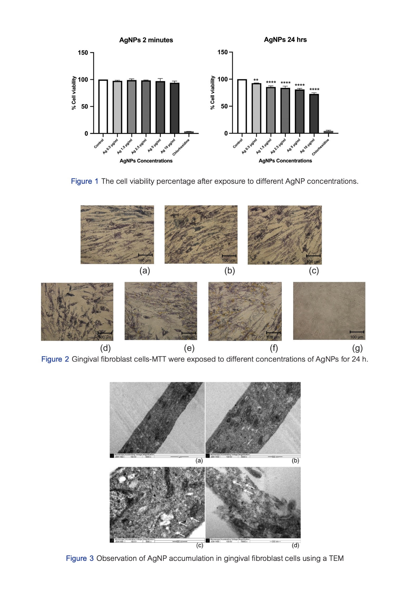
Results: A 2-min exposure to 0.5−10 μg/mL AgNPs did not induce cytotoxicity. In contrast, a 24-h exposure led to a significant decrease in cell viability, however, it remained above 70% compared with the control. Exposure to AgNPs for 24 h resulted in a lower density of HGF cells and more degenerative cells when the concentration of AgNPs increased observed with the microscope. TEM analysis revealed the absorption, internalization, and dissemination of 0.5 μg/mL AgNPs in HGF cells after a 24-h incubation.
Conclusions: The cytotoxicity of AgNPs was concentration- and exposure time-dependant, as evidenced by the intracellular accumulation observed using TEM after a 24-h exposure. Concentrations below 10 μg/mL can be considered non-cytotoxic during short-term oral cavity exposure. Based on our results, the use of AgNPs in dentistry is likely to be safe for oral application.
Article Details

This work is licensed under a Creative Commons Attribution-NonCommercial-NoDerivatives 4.0 International License.
References
Corrêa JM, Mori M, Sanches HL, da Cruz AD, Poiate E Jr, Poiate IA. Silver nanoparticles in dental biomaterials. Int J Biomater. 2015;2015:485275. doi: 10.1155/2015/485275.
Noronha VT, Paula AJ, Durán G, Galembeck A, Cogo-Müller K, Franz-Montan M, et al. Silver nanoparticles in dentistry. Dent Mater. 2017 Oct;33(10):1110-1126. doi: 10.1016/j.dental.2017.07.002.
Inkielewicz-Stepniak I, Santos-Martinez MJ, Medina C, Radomski MW. Pharmacological and toxicological effects of co-exposure of human gingival fibroblasts to silver nanoparticles and sodium fluoride. Int J Nanomedicine 2014 Apr;9:1677-1687. doi: 10.2147/IJN.S59172.
Freire PL, Stamford TC, Albuquerque AJ, Sampaio FC, Cavalcante HM, Macedo RO, et al. Action of silver nanoparticles towards biological systems: cytotoxicity evaluation using hen's egg test and inhibition of Streptococcus mutans biofilm formation. Int J Antimicrob Agents. 2015 Feb;45(2):183-187. doi: 10.1016/j.ijantimicag.2014.09.007.
Haghgoo R, Saderi H, Eskandari M, Haghshenas H, Rezvani M. Evaluation of the antimicrobial effect of conventional and nanosilver-containing varnishes on oral streptococci. J Dent. (Shiraz) 2014 Jun;15(2):57-62.
Pérez-Díaz MA, Boegli L, James G, Velasquillo C, Sánchez-Sánchez R, Martínez-Martínez RE, et al. Silver nanoparticles with antimicrobial activities against Streptococcus mutans and their cytotoxic effect. Mater Sci Eng C Mater Biol Appl. 2015 Oct;55:360-366. doi: 10.1016/j.msec.2015.05.036.
Iravani S, Korbekandi H, Mirmohammadi SV, Zolfaghari B. Synthesis of silver nanoparticles: chemical, physical and biological methods. Res Pharm Sci. 2014 Nov-Dec;9(6):385-406.
Santos VE Jr, Vasconcelos Filho A, Targino AG, Flores MA, Galembeck A, Caldas AF Jr, et al. A new "silver-bullet" to treat caries in children--nano silver fluoride: a randomised clinical trial. J Dent. 2014 Aug;42(8):945-951. doi: 10.1016/j.jdent.2014.05.017.
Akter M, Sikder MT, Rahman MM, Ullah AKMA, Hossain KFB, Banik S, et al. A systematic review on silver nanoparticles-induced cytotoxicity: Physicochemical properties and perspectives. J Adv Res. 2017 Nov;9:1-16. doi: 10.1016/j.jare.2017.10.008.
AshaRani PV, Low Kah Mun G, Hande MP, Valiyaveettil S. Cytotoxicity and genotoxicity of silver nanoparticles in human cells. ACS Nano. 2009 Feb;3(2):279-290. doi: 10.1021/nn800596w.
Taleghani F, Yaraii R, Sadeghi R, Haghgoo R, Rezvani MB. Cytotoxicity of silver nanoparticles on human gingival epithelial cells: An in vitro study. J Dent Sch. 2019 Mar;32(1):30-36. doi: 10.22037/jds.v32i1.24760.
Pisitpongthon N. In vitro study on the effectiveness of nanosilver fluoride in biofilm formation inhibition and biofilm susceptibility. Thesis, Mahidol University 2020.
Nagireddy VR, Reddy D, Kondamadugu S, Puppala N, Mareddy A, Chris A. Nanosilver fluoride-a paradigm shift for arrest in dental caries in primary teeth of school children: A Randomized Controlled Clinical Trial. Int J Clin Pediatr Dent. 2019 Nov-Dec;12(6):484-490. doi: 10.5005/jp-journals-10005-1703.
Al-Nerabieah Z, Arrag E, Rajab A. Cariostatic efficacy and children acceptance of nano-silver fluoride versus silver diamine fluoride: a randomized controlled clinical trial. J Stomatol. 2020;73(3):100-106. doi: 10.5114/jos.2020.96939.
Wu M, Guo H, Liu L, Liu Y, Xie L. Size-dependent cellular uptake and localization profiles of silver nanoparticles. Int J Nanomedicine 2019;14:4247-4259. doi: 10.2147/IJN.S201107.
Kang B, Mackey MA, El-Sayed MA. Nuclear targeting of gold nanoparticles in cancer cells induces DNA damage, causing cytokinesis arrest and apoptosis. J Am Chem Soc. 2010;132(5):1517-1519. doi: 10.1021/ja9102698.
Dawson KA, Salvati A, Lynch I. Nanotoxicology: nanoparticles reconstruct lipids. Nat Nanotechnol. 2009 Feb;4(2):84-85. doi: 10.1038/nnano.2008.426.
Wakshlak RB, Pedahzur R, Avnir D. Antibacterial activity of silver-killed bacteria: the "zombies" effect. Sci Rep. 2015 Apr;5:9555. doi: 10.1038/srep09555.
Haase A, Rott S, Mantion A, Graf P, Plendl J, Thünemann AF, et al. Effects of silver nanoparticles on primary mixed neural cell cultures: uptake, oxidative stress and acute calcium responses. Toxicol Sci. 2012 Apr;126(2):457-468. doi: 10.1093/toxsci/kfs003.
Riaz Ahmed KB, Nagy AM, Brown RP, Zhang Q, Malghan SG, Goering PL. Silver nanoparticles: Significance of physicochemical properties and assay interference on the interpretation of in vitro cytotoxicity studies. Toxicol In Vitro. 2017 Feb;38:179-192. doi: 10.1016/j.tiv.2016.10.012.
Hsiao IL, Hsieh YK, Wang CF, Chen IC, Huang YJ. Trojan-horse mechanism in the cellular uptake of silver nanoparticles verified by direct intra- and extracellular silver speciation analysis. Environ Sci Technol. 2015 Mar;49(6):3813-3821. doi: 10.1021/es504705p.
Arora S, Jain J, Rajwade JM, Paknikar KM. Interactions of silver nanoparticles with primary mouse fibroblasts and liver cells. Toxicol Appl Pharmacol. 2009 May;236(3):310-318. doi: 10.1016/j.taap.2009.02.020.
Ahamed M, Karns M, Goodson M, Rowe J, Hussain SM, Schlager JJ, et al. DNA damage response to different surface chemistry of silver nanoparticles in mammalian cells. Toxicol Appl Pharmacol. 2008 Dec;233(3):404-410. doi: 10.1016/j.taap.2008.09.015.
Zhang T, Wang L, Chen Q, Chen C. Cytotoxic potential of silver nanoparticles. Yonsei Med J. 2014 Mar;55(2):283-291. doi: 10.3349/ymj.2014.55.2.283.
Heshmati M, Arbabi Bidgoli S, Khoei S, Mahmoudzadeh A, Sorkhabadi S. Cytotoxicity and genotoxicity of silver nanoparticles in Chinese Hamster ovary cell line (CHO-K1) cells. The Nucleus 2019;62. doi:10.1007/s13237-019-00295-y.
Chernousova S, Epple M. Silver as antibacterial agent: ion, nanoparticle, and metal. Angew Chem Int Ed Engl. 2013;52(6):1636-1653. doi: 10.1002/anie.201205923.


