
Characteristics and osteoblast cell responses of the anodized titanium and sandblasted and acid-etched titanium surfaces
Article Sidebar
Main Article Content
Abstract
Objectives: The purpose of this article was to study the topographic properties and cellular responses of the anodized titanium surface and sandblasted and acid-etched surface.
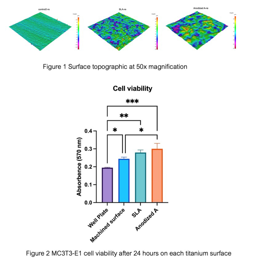
Material and Methods: Nine titanium discs were fabricated and treated with 3 types of titanium surface treatment (3-machined surface, 3-sandblasted and acid-etched surface, and 3-anodized surface). Each titanium surface was tested with profilometer to measure the surface roughness and captured 3D profile images. MC3T3-E1 cells were seeded onto titanium disc to test the cell viability. Cells were seeded on each surface treatment at 4 hours and 6 hours, fixed with glutaraldehyde solution, dehydrated, and then gold coated to study surface morphology with scanning electron microscope image.
Results: The anodized titanium surface and sandblasted and acid-etched surface exhibited more favorable topographical characteristics compared to the machined surface (control group), with an average roughness of 1.34, and 1.05 μm respectively. The spreading of osteoblast-like cells on the anodized titanium surface was similar to the sandblasted and acid-etched surfaces. While the spreading of osteoblast-like cells on the anodized titanium surface was observed more cytoplasmic projections and cell size than the cell observed in the machined surface. Cellular viability showed a statistically significant difference between the anodized and machined surface (p<0.05). No statistically significant difference was found between the anodized and sandblasted
and acid-etched surface (p<0.05).
Conclusions: The topography and the spreading of osteoblast-like cells on the anodized titanium surface exhibit statistically significant differences compared to the machined surface. However, there is no statistically significant difference in the spreading of osteoblast-like cells between the anodized titanium surface and the sandblasted and acid-etched surface.
Article Details

This work is licensed under a Creative Commons Attribution-NonCommercial-NoDerivatives 4.0 International License.
References
Parithimarkalaignan S, Padmanabhan TV. Osseointegration: an update. J Indian Prosthodont Soc. 2013 Mar;13(1):2-6. doi:10.1007/s13191-013-0252-z.
Pandey C, Rokaya D, Bhattarai BP. Contemporary concepts in osseointegration of Dental Implants: A Review. Biomed Res Int. 2022 Jun;2022:6170452. doi:10.1155/2022/6170452.
Wennerberg A, Albrektsson T. On implant surfaces: a review of current knowledge and opinions. Int J Oral Maxillofac Implants. 2010 Jan-Feb;25(1):63-74.
Puleo DA, Thomas MV. Implant surfaces. Dent Clin North Am. 2006 Jul;50(3):323-338, v. doi:10.1016/j.cden.2006.03.001.
Li D, Ferguson SJ, Beutler T, Cochran DL, Sittig C, Hirt HP, et al. Biomechanical comparison of the sandblasted and acid-etched and the machined and acid-etched titanium surface for dental implants. J Biomed Mater Res. 2002 May;60(2):325-332. doi:10.1002/jbm.10063.
Cochran DL, Buser D, ten Bruggenkate CM, Weingart D, Taylor TM, Bernard JP, et al. The use of reduced healing times on ITI implants with a sandblasted and acid-etched (SLA) surface: early results from clinical trials on ITI SLA implants. Clin Oral Implants Res. 2002 Apr;13(2):144-153. doi:10.1034/j.1600-0501.2002.130204.x.
Su EP, Justin DF, Pratt CR, Sarin VK, Nguyen VS, Oh S, et al. Effects of titanium nanotubes on the osseointegration, cell differentiation, mineralisation and antibacterial properties of orthopaedic implant surfaces. Bone Joint J. 2018 Jan;100-B(1 Supple A):9-16. doi:10.1302/0301-620X.100B1.BJJ-2017-0551.R1
Inchingolo AM, Malcangi G, Ferrante L, Del Vecchio G, Viapiano F, Inchingolo AD, et al. Surface coatings of dental implants: A review. J Funct Biomater. 2023 May 22;14(5):287. doi:10.3390/jfb14050287.
Lavenus S, Louarn G, Layrolle P. Nanotechnology and dental implants. Int J Biomater. 2010;2010:915327. doi:10.1155/2010/915327
Sykaras N, Iacopino AM, Marker VA, Triplett RG, Woody RD. Implant materials, designs, and surface topographies: their effect on osseointegration. A literature review. Int J Oral Maxillofac Implants. 2000 Sep-Oct;15(5):675-690.
Sharma A, McQuillan AJ, Sharma LA, Waddell JN, Shibata Y, Duncan WJ. Spark anodization of titanium-zirconium alloy: surface characterization and bioactivity assessment. J Mater Sci Mater Med. 2015 Aug;26(8):221. doi:10.1007/s10856-015-5555-7.
Ivanoff CJ, Widmark G, Johansson C, Wennerberg A. Histologic evaluation of bone response to oxidized and turned titanium micro-implants in human jawbone. Int J Oral Maxillofac Implants. 2003 May-Jun;18(3):341-348.
Jungner M, Lundqvist P, Lundgren S. Oxidized titanium implants (Nobel Biocare TiUnite) compared with turned titanium implants (Nobel Biocare mark III) with respect to implant failure in a group of consecutive patients treated with early functional loading and two-stage protocol. Clin Oral Implants Res. 2005 Jun;16(3):308-312. doi:10.1111/j.1600-0501.2005.01101.x.
Wennerberg A, Albrektsson T. Suggested guidelines for the topographic evaluation of implant surfaces. Int J Oral Maxillofac Implants. 2000 May-Jun;15(3):331-344.
Zhu X, Chen J, Scheideler L, Reichl R, Geis-Gerstorfer J. Effects of topography and composition of titanium surface oxides on osteoblast responses. Biomaterials. 2004 Aug;25(18):4087-4103. doi:10.1016/j.biomaterials.2003.11.011.
Eriksson C, Lausmaa J, Nygren H. Interactions between human whole blood and modified TiO2-surfaces: influence of surface topography and oxide thickness on leukocyte adhesion and activation. Biomaterials. 2001 Jul;22(14):1987-1996. doi:10.1016/s0142-9612(00)00382-3.
Martin JY, Schwartz Z, Hummert TW, Schraub DM, Simpson J, Lankford J Jr, et al. Effect of titanium surface roughness on proliferation, differentiation, and protein synthesis of human osteoblast-like cells (MG63). J Biomed Mater Res. 1995 Mar;29(3):389-401. doi:10.1002/jbm.820290314.
Sul YT, Johansson CB, Jeong Y, Wennerberg A, Albrektsson T. Resonance frequency and removal torque analysis of implants with turned and anodized surface oxides. Clin Oral Implants Res. 2002 Jun;13(3):252-259. doi:10.1034/j.1600-0501.2002.130304.x
De Angelis E, Ravanetti F, Cacchioli A, Corradi A, Giordano C, Candiani G, et al. Attachment, proliferation and osteogenic response of osteoblast-like cells cultured on titanium treated by a novel multiphase anodic spark deposition process. J Biomed Mater Res B Appl Biomater. 2009 Jan;88(1):280-289. doi:10.1002/jbm.b.31179.
Kim MH, Park K, Choi KH, Kim SH, Kim SE, Jeong CM, et al. Cell adhesion and in vivo osseointegration of sandblasted/acid etched/anodized dental implants. Int J Mol Sci. 2015 May;16(5):10324-10336. doi:10.3390/ijms160510324.
Sista S, Nouri A, Li Y, Wen C, Hodgson PD, Pande G. Cell biological responses of osteoblasts on anodized nanotubular surface of a titanium-zirconium alloy. J Biomed Mater Res A. 2013 Dec;101(12):3416-3430. doi:10.1002/jbm.a.34638.
Zhao G, Schwartz Z, Wieland M, Rupp F, Geis-Gerstorfer J, Cochran DL, et al. High surface energy enhances cell response to titanium substrate microstructure. J Biomed Mater Res A. 2005 Jul 1;74(1):49-58. doi:10.1002/jbm.a.30320
Fabbro MD, Taschieri S, Canciani E, Addis A, Musto F, Weinstein R, et al. Osseointegration of titanium implants with different rough surfaces: A histologic and histomorphometric study in an adult minipig model. Implant Dent. 2017 Jun;26(3):357-366. doi:10.1097/ID.0000000000000560.
De Maeztu MA, Braceras I, Alava JI, Sánchez-Garcés MA, Gay-Escoda C. Histomorphometric study of ion implantation and diamond-like carbon as dental implant surface treatments in beagle dogs. Int J Oral Maxillofac Implants. 2007 Mar-Apr;22(2):273-279.
