pH changes of root dentin following root canal filling with three different calcium silicate-based cements over 4-week period
Main Article Content
Abstract
Objective: To evaluate the pH changes in root dentin after root canals were filled with ProRoot MTA, Bio-MA or Biodentine at different root levels and times.
Materials and Methods: Fifty-three extracted single-rooted mandibular premolars were instrumented, and three different locations of cavities were prepared on the root surface-two at the middle and one at the apical root levels. Three experimental groups (n=16) were filled with ProRoot MTA, Bio-MA or Biodentine, while a control group (n=5) was filled with deionized water. pH measurements were taken at 2 hours, 1 day, 1-4 weeks. Analysis was performed using the three-way mixed ANOVA (p< 0.05).
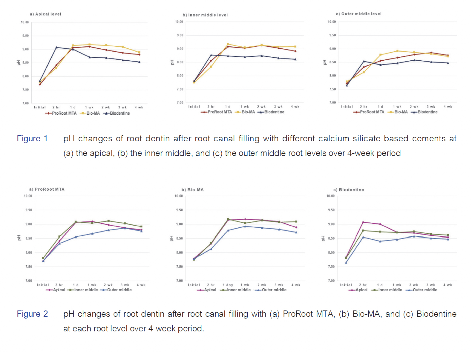
Results: Significant alkaline pH changes occurred over a 4-week period in experimental groups at all root levels (p < .001). The Bio-MA group exhibited the highest pH value (9.18) at 1 week at the apical root level and 1 day at the inner middle root levels. The highest pH values for ProRoot MTA and Biodentine groups were 9.12 and 9.07 at 2 weeks and 2 hours at the inner middle and apical root levels, respectively. Notably, the pH values at the outer middle root level were the lowest.
Conclusions: Alkaline pH changes in root dentin persisted for 4 weeks, with the highest recorded values ranging from 9.07-9.18. The outer middle root level displayed the lowest pH values.
Article Details

This work is licensed under a Creative Commons Attribution-NonCommercial-NoDerivatives 4.0 International License.
References
Karamifar K, Tondari A, Saghiri MA. Endodontic periapical lesion: An overview on the etiology, diagnosis and current treatment Modalities. Eur Endod J. 2020 Jul;5(2):54-67. doi: 10.14744/eej.2020.42714.
Baugh D, Wallace J. The role of apical instrumentation in root canal treatment: a review of the literature. J Endod 2005 May;31(5):333-340. doi:10.1097/01.don.0000145422.94578.e6.
Trope M, Bunes A, Debelian G. Root filling materials and techniques: bioceramics a new hope?. Endod Topics 2015 Jan;32(1):86-96. doi: 10.1111/etp.12074.
Ørstavik D. Materials used for root canal obturation: technical, biological and clinical testing. Endod Topics 2005 Jan;12(1):25-38. doi.org/10.1111/j.1601-1546.2005.00197.x.
Bogen G, Kuttler S. Mineral trioxide aggregate obturation: a review and case series. J Endod 2009 Jun; 35(6):777-790. doi: 10.1016/j.joen.2009.03.006.
Eskandari F, Razavian A, Hamidi R, Yousefi K, Borzou S. An updated review on properties and indications of calcium silicate-based cements in endodontic therapy. Int J Dent 2022 Oct; 2022:6858088. doi:10.1155/2022/6858088.
Parirokh M, Torabinejad M. Mineral trioxide aggregate: a comprehensive literature review-Part III: Clinical applications, drawbacks, and mechanism of action. J Endod 2010 Mar;36(3):400-413. doi:10.1016/j.joen.2009.09.009.
Camilleri J. Characterization of hydration products of mineral trioxide aggregate. Int Endod J 2008 May;41(5):408-417. doi: 10.1111/j.1365-2591.2007.01370.x.
Parirokh M, Torabinejad M. Mineral trioxide aggregate: a comprehensive literature review-Part I: chemical, physical, and antibacterial properties. J Endod 2010 Jan;36(1):16-27. doi: 10.1016/j.joen.2009.09.006.
Al-Hezaimi K, Al-Shalan TA, Naghshbandi J, Oglesby S, Simon JH, Rotstein I. Antibacterial effect of two mineral trioxide aggregate (MTA) preparations against Enterococcus faecalis and Streptococcus sanguis in vitro. J Endod 2006 Nov;32(11):1053-1056. doi: 10.1016/j.joen.2006.06.004.
Nerwich A, Figdor D, Messer HH. pH changes in root dentin over a 4-week period following root canal dressing with calcium hydroxide. J Endod 1993 Jun;19(6):302-306. doi: 10.1016/s0099-2399(06)80461-9.
Teixeira FB, Levin LG, Trope M. Investigation of pH at different dentinal sites after placement of calcium hydroxide dressing by two methods. Oral Surg Oral Med Oral Pathol Oral Radiol Endod 2005 Apr;99(4):511-516. doi: 10.1016/j.tripleo.2004.07.023.
Camargo CH, Bernardineli N, Valera MC, de Carvalho CA, de Oliveira LD, Menezes MM, et al. Vehicle influence on calcium hydroxide pastes diffusion in human and bovine teeth. Dent Traumatol 2006 Dec;22(6):302-306. doi: 10.1111/j.1600-9657.2005.00326.x.
Chamberlain TM, Kirkpatrick TC, Rutledge RE. pH changes in external root surface cavities after calcium hydroxide is placed at 1, 3 and 5 mm short of the radiographic apex. Dent Traumatol 2009 Oct;25(5):470-474. doi: 10.1111/j.1600-9657.2009.00806.x.
Heward S, Sedgley CM. Effects of intracanal mineral trioxide aggregate and calcium hydroxide during four weeks on pH changes in simulated root surface resorption defects: an in vitro study using matched pairs of human teeth. J Endod 2011 Jan;37(1):40-44. doi: 10.1016/j.joen.2010.09.003.
Hansen SW, Marshall JG, Sedgley CM. Comparison of intracanal EndoSequence Root Repair Material and ProRoot MTA to induce pH changes in simulated root resorption defects over 4 weeks in matched pairs of human teeth. J Endod 2011 Apr;37(4):502-506. doi: 10.1016/j.joen.2011.01.010.
Ozok AR, Wu MK, Wesselink PR. Comparison of the in vitro permeability of human dentine according to the dentinal region and the composition of the simulated dentinal fluid. J Dent 2002 Feb-Mar;30(2-3):107-111. doi: 10.1016/s0300-5712(02)00005-2.
Kaur M, Singh H, Dhillon JS, Batra M, Saini M. MTA versus Biodentine: review of literature with a comparative analysis. J Clin Diagn Res 2017 Aug;11(8): ZG01-ZG05. doi: 10.7860/JCDR/2017/25840.10374.
Trongkij P, Sutimuntanakul S, Lapthanasupkul P, Chaimanakarn C, Wong R, Banomyong D. Effects of the exposure site on histological pulpal responses after direct capping with 2 calcium-silicate based cements in a rat model. Restor Dent Endod 2018 Aug;43(4):e36. doi: 10.5395/rde.2018.43.e36.
Torabinejad M, Hong CU, McDonald F, Pitt Ford TR. Physical and chemical properties of a new root-end filling material. J Endod 1995 Jul;21(7): 349-353. doi: 10.1016/S0099-2399(06)80967-2.
Ozdemir HO, Ozçelik B, Karabucak B, Cehreli ZC. Calcium ion diffusion from mineral trioxide aggregate through simulated root resorption defects. Dent Traumatol 2008 Feb;24(1) 70-73. doi: 10.1111/j.1600-9657.2006.00512.x.
Warotamawichaya S, Sutimuntanakul S. Effect of calcium chloride on setting time of Thai white Portland cement [High Grad Dip Project Report]. Bangkok: Mahidol University; 2011.
Malkondu Ö, Karapinar Kazandağ M, Kazazoğlu E. A review on biodentine, a contemporary dentine replacement and repair material. Biomed Res Int 2014;2014:160951. doi:10.1155/2014/160951.
Camilleri J. Hydration mechanisms of mineral trioxide aggregate. Int Endod J 2007 Jun;40(6): 462-470. doi: 10.1111/j.1365-2591.2007.01248.x.
Islam I, Chng HK, Yap AU. Comparison of the physical and mechanical properties of MTA and Portland cement. J Endod 2006 Mar;32(3): 193-197. doi: 10.1016/j.joen.2005.10.043.
Lee YL, Lee BS, Lin FH, Yun Lin A, Lan WH, Lin CP. Effects of physiological environments on the hydration behavior of mineral trioxide aggregate. Biomaterials 2004 Feb;25(5):787-793. doi: 10.1016/s0142-9612(03)00591-x.
Fridland M, Rosado R. Mineral trioxide aggregate (MTA) solubility and porosity with different water-to-powder ratios. J Endod 2003 Dec;29(12):814-817. doi: 10.1097/00004770-200312000-00007.
Talabani RM, Garib BT, Masaeli R. Bioactivity and physicochemical properties of three calcium silicate-based cements: An in vitro study. Biomed Res Int 2020 May;2020:9576930 doi:10.1155/2020/9576930.
Zeid STA, Alothmani OS, Yousef MK. Biodentine and mineral trioxide aggregate: an analysis of solubility, pH changes and leaching elements. Life Sci J 2015 Apr;12(4):18- 23.
Quintana RM, Jardine AP, Grechi TR, Grazziotin-Soares R, Ardenghi DM, Scarparo RK, et al. Bone tissue reaction, setting time, solubility, and pH of root repair materials. Clin Oral Investig 2019 Mar;23(3):1359-1366. doi: 10.1007/s00784-018-2564-1.
Grech L, Mallia B, Camilleri J. Characterization of set Intermediate Restorative Material, Biodentine, Bioaggregate and a prototype calcium silicate cement for use as root‐end filling materials. Int Endod J 2013 Jul;46(7): 632-641. doi: 10.1111/iej.12039.
Carrigan PJ, Morse DR, Furst ML, Sinai IH. A scanning electron microscopic evaluation of human dentinal tubules according to age and location. J Endod 1984 Aug;10(8):359-363. doi: 10.1016/S0099-2399(84)80155-7.
Marion D, Jean A, Hamel H, Kerebel LM, Kerebel B. Scanning electron microscopic study of odontoblasts and circumpulpal dentin in a human tooth. Oral Surg Oral Med Oral Pathol Oral Radiol Endod 1991 Oct;72(4): 473-478. doi: 10.1016/0030-4220(91)90563-r.
Tidmarsh BG, Arrowsmith MG. Dentinal tubules at the root ends of apicected teeth: a scanning electron microscopic study. Int Endod J 1989 Jul;22(4):184-189. doi: 10.1111/j.1365-2591.1989.tb00922.x.
Takahashi N, Schachtele CF. Effect of pH on the growth and proteolytic activity of Porphyromonas gingivalis and Bacteroides intermedius. J Dent Res 1990 Jun;69(6):1266-1269. doi: 10.1177/00220345900690060801.
Takahashi N, Saito K, Schachtele CF, Yamada T. Acid tolerance and acid‐neutralizing activity of Porphyromonas gingivalis, Prevotella intermedia and Fusobacterium nucleatum. Oral Microbial Immunol 1997 Dec;12(6):323-328. doi: 10.1111/j.1399-302x.1997.tb00733.x.
Siqueira JF Jr, Lopes HP. Mechanisms of antimicrobial activity of calcium hydroxide: a critical review. Int Endod J 1999 Sep;32(5):361-369. doi: 10.1046/j.1365-2591.1999.00275.x.
Trope M, Moshonov J, Nissan R, Buxt P, Yesilsoy C. Short vs. long‐term calcium hydroxide treatment of established inflammatory root resorption in replanted dog teeth. Endod Dent Traumatol 1995 Jun;11(3):124-128. doi: 10.1111/j.1600-9657.1995.tb00473.x.
Aggarwal V, Singla M. Management of inflammatory root resorption using MTA obturation– a four year follow up. Br Dent J 2010 Apr;208(7):287-289. doi: 10.1038/sj.bdj.2010.293.
Asgary S, Nosrat A, Seifi A. Management of inflammatory external root resorption by using calcium-enriched mixture cement: a case report. J Endod 2011 Mar;37(3):411-413. doi: 10.1016/j.joen.2010.11.015.


