
In vitro cytotoxicity evaluation of an experimental orthodontic adhesive containing gold nanoparticles
Article Sidebar
Main Article Content
Abstract
Objective: This study aimed to evaluate and compare the in vitro cytotoxicity of an experimental orthodontic adhesive containing 0.5% wt gold nanoparticles (AuNPs) with a conventional light-cured adhesive (Transbond XT) on human gingival fibroblast (HGF) cells using the MTT assay.
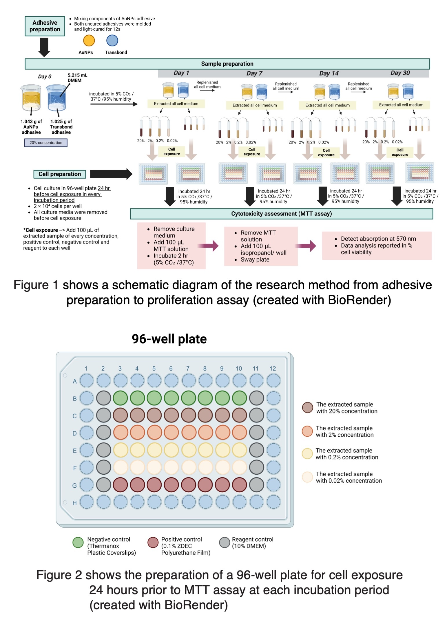
Materials and Methods: Both adhesives, comprising the AuNPs group and the Transbond group, were prepared and incubated in Dulbecco’s Modified Eagle Medium (DMEM) under the same conditions with 20% concentration for 1, 7, 14, and 30 days. After each incubation, the whole medium was extracted for analysis, and the fresh medium was replenished at the same amount. Extracts were tested at concentrations of 20%, 2%, 0.2%, and 0.02%. HGF cells were seeded in 96-well plates 24 hours before cell exposure at each incubation and performing MTT assays. Cell viability was measured spectrophotometrically and analyzed using independent sample t-tests (p<0.05).
Results: At a 20% concentration, the AuNPs group exhibited significantly lower cell viability than the Transbond group across all time points, with severe cytotoxicity observed on Days 1 and 7, moderate on Day 14, and mild on Day 30. In contrast, the Transbond group consistently showed mild cytotoxicity. Both groups showed no cytotoxicity at lower concentrations (2%, 0.2%, 0.02%). Interestingly, at 2% and 0.2% concentrations, the AuNPs group had significantly higher cell viability than the Transbond group in most periods. A general trend of decreasing cytotoxicity over time was observed for both adhesives.
Conclusions: AuNPs adhesive showed higher cytotoxicity than the conventional adhesive at undiluted extract (20%) and both materials were non-cytotoxic at lower concentrations. These findings highlight the importance of concentration and aging in cytotoxicity outcomes and suggest that AuNPs adhesive may be biocompatible under clinical conditions. Further, in vivo studies are necessary to confirm the safety of AuNPs adhesive.
Article Details

This work is licensed under a Creative Commons Attribution-NonCommercial-NoDerivatives 4.0 International License.
References
Khoroushi M, Kachuie M. Prevention and treatment of white spot lesions in orthodontic patients. Contemp Clin Dent. 2017 Jan-Mar;8(1):11-19. doi: 10.4103/ccd.ccd_216_17
Cheng L, Zhang K, Weir MD, Melo MA, Zhou X, Xu HH. Nanotechnology strategies for antibacterial and remineralizing composites and adhesives to tackle dental caries. Nanomedicine (Lond). 2015 Mar;10(4):627-641. doi: 10.2217/nnm.14.191
Lalit H, Subramanian A, Sivashanmugam P. Preparation, characterization, and evaluation of cytotoxic activity of a novel titanium dioxide nanoparticle-infiltrated orthodontic adhesive: An In vitro study. World J Dent. 2023 Nov;14:882-887. doi: 10.5005/jp-journals-10015-2319
Dechsakulthorn F, Hayes A, Bakand S, Joeng L, Winder C. In vitro cytotoxicity assessment of selected nanoparticles using human skin fibroblasts. AATEX 2006 Nov; 14:397-400.
Lanone S, Rogerieux F, Geys J, Dupont A, Maillot-Marechal E, Boczkowski J, et al. Comparative toxicity of 24 manufactured nanoparticles in human alveolar epithelial and macrophage cell lines. Part Fibre Toxicol. 2009 Apr 30;6:14. doi: 10.1186/1743-8977-6-14
Wang B, Feng WY, Wang TC, Jia G, Wang M, Shi JW, et al. Acute toxicity of nano- and micro-scale zinc powder in healthy adult mice. Toxicol Lett. 2006 Feb 20;161(2):115-123. doi: 10.1016/j.toxlet.2005.08.007
Cabuzu D, Cirja A, Puiu R, Grumezescu AM. Biomedical applications of gold nanoparticles. Curr Top Med Chem. 2015;15(16):1605-1613. doi: 10.2174/1568026615666150414144750
Akarajarasrod P, Dechkunakorn S, Tantivitayakul P, Punnakitikashem P, Wichai W, Whitis PP, et al. Antibacterial effect of experimental orthodontic adhesives containing gold nanoparticles against Streptococcus mutans and Streptococcus sobrinus. Key Eng Mater. 2021 Nov;904:301-308. doi: 10.4028/www.scientific.net/KEM.904.301
Kus-Liśkiewicz M, Fickers P, Ben Tahar I. Biocompatibility and cytotoxicity of gold nanoparticles: Recent advances in methodologies and regulations. Int J Mol Sci. 2021 Oct 11;22(20):10952. doi: 10.3390/ijms222010952
Huang TH, Tsai CY, Chen SL, Kao CT. An evaluation of the cytotoxic effects of orthodontic bonding adhesives upon a primary human oral gingival fibroblast culture and a permanent, human oral cancer-cell line. J Biomed Mater Res. 2002;63(6):814-821. doi: 10.1002/jbm.10412
Heravi F, Ramezani M, Poosti M, Hosseini M, Shajiei A, Ahrari F. In vitro cytotoxicity assessment of an orthodontic composite containing titanium-dioxide nano-particles. J Dent Res Dent Clin Dent Prospects. 2013 Dec;7(4):192-198. doi: 10.5681/joddd.2013.031
Malkoc S, Corekci B, Ulker HE, Yalçin M, Sengün A. Cytotoxic effects of orthodontic composites. Angle Orthod. 2010 Jul;80(4):571-576. doi: 10.2319/092809-537.1
Wozniak A, Malankowska A, Nowaczyk G, Grześkowiak B, Tuśnio K, Slomski R, et al. Size and shape-dependent cytotoxicity profile of gold nanoparticles for biomedical applications. J Mater Sci Mater Med. 2017 Jun;28(6):92. doi: 10.1007/s10856-017-5902-y
Nimcharoensuk K, Anuwongnukroh N, Dechkunakorn S, Sattabanasuk V, Wichai W, Sunintaboon P. Shear bond strength of experimental light-cured orthodontic adhesives. Key Eng Mater. 2019 Jul;814:378-383. doi: 10.4028/www.scientific.net/KEM.814.378
International Organization for Standardization Biological evaluation of medical devices. Part 12: Sample preparation and reference materials (ISO standard no.10993-12:2021). 2021.
International Organization for Standardization Biological evaluation of medical devices. Part 5: Tests for in vitro cytotoxicity (ISO standard no.10993-5:2009 (E)). 2009.
Sjögren G, Sletten G, Dahl JE. Cytotoxicity of dental alloys, metals, and ceramics assessed by millipore filter, agar overlay, and MTT tests. J Prosthet Dent. 2000 Aug;84(2):229-236. doi: 10.1067/mpr.2000.107227
Dahl JE, Frangou-Polyzois MJ, Polyzois GL. In vitro biocompatibility of denture relining materials. Gerodontology. 2006 Mar;23(1):17-22. doi: 10.1111/j.1741-2358.2006.00103.x
Bationo R, Rouamba A, Diarra A, Beugré-Kouassi MLA, Beugré JB, Jordana F. Cytotoxicity evaluation of dental and orthodontic light-cured composite resins. Clin Exp Dent Res. 2021 Feb;7(1):40-48. doi: 10.1002/cre2.337
Ferracane JL. Elution of leachable components from composites. J Oral Rehabil. 1994 Jul;21(4):441-452. doi: 10.1111/j.1365-2842.1994.tb01158.x
Thompson LR, Miller EG, Bowles WH. Leaching of unpolymerized materials from orthodontic bonding resin. J Dent Res. 1982 Aug;61(8):989-992. doi: 10.1177/00220345820610081501
Ahrari F, Tavakkol Afshari J, Poosti M, Brook A. Cytotoxicity of orthodontic bonding adhesive resins on human oral fibroblasts. Eur J Orthod. 2010 Dec;32(6):688-692. doi: 10.1093/ejo/cjq019
Goldberg M. In vitro and in vivo studies on the toxicity of dental resin components: a review. Clin Oral Investig. 2008 Mar;12(1):1-8. doi: 10.1007/s00784-007-0162-8
Gupta SK, Saxena P, Pant VA, Pant AB. Release and toxicity of dental resin composite. Toxicol Int. 2012 Sep;19(3):225-234. doi: 10.4103/0971-6580.103652
Gorgen V, Guler C. Residual monomer in dentistry: A literature review [Dis hekimliginde artik monomerler: Bir liteartur derlemesi]. Med Sci. 2015;4(1):2024 - 2038. doi: 10.5455/medscience.2014.03.8200
Odabasi D, Guler C, Kucukaslan D. Evaluation of the amount of residual monomer released from different flowable composite resins. BMC Oral Health. 2024 Feb 15;24(1):244. doi: 10.1186/s12903-024-04005-2
Solanki LA, Dinesh SPS, Jain RK, Balasubramaniam A. Effects of titanium oxide coating on the antimicrobial properties, surface characteristics, and cytotoxicity of orthodontic brackets - A systematic review and meta analysis of in-vitro studies. J Oral Biol Craniofac Res. 2023 Sep-Oct;13(5):553-562. doi: 10.1016/j.jobcr.2023.05.014
Jia Y, Shi K, Liao J, Peng J, Hao Y, Qu Y, et al. Effects of cetyltrimethylammonium bromide on the toxicity of gold nanorods both in vitro and in vivo: molecular origin of cytotoxicity and Inflammation. Small Methods. 2020;4:1900799. doi: 10.1002/smtd.201900799
Mohsen NM, Craig RG, Hanks CT. Cytotoxicity of urethane dimethacrylate composites before and after aging and leaching. J Biomed Mater Res. 1998 Feb;39(2):252-260. doi: 10.1002/(sici)1097-4636(199802)39:2<252::aid-jbm12>3.0.co;2-f
Franz A, König F, Lucas T, Watts DC, Schedle A. Cytotoxic effects of dental bonding substances as a function of degree of conversion. Dent Mater. 2009 Feb;25(2):232-239. doi: 10.1016/j.dental.2008.07.003
Jagdish N, Padmanabhan S, Chitharanjan AB, Revathi J, Palani G, Sambasivam M, et al. Cytotoxicity and degree of conversion of orthodontic adhesives. Angle Orthod. 2009 Nov;79(6):1133-1138. doi: 10.2319/080808-418r.1
Samuelsen JT, Dahl JE. Biological aspects of modern dental composites. Biomater Investig Dent. 2023 Jun 19;10(1):2223223. doi: 10.1080/26415275.2023.2223223
Alizadehgharib S, Östberg AK, Dahlgren U. Effects of the methacrylate/acrylate monomers HEMA, TEGDMA, DEGDA, and EMA on the immune system. Clin Exp Dent Res. 2017 Nov 17;3(6):227-234. doi: 10.1002/cre2.93
Klenotová M, Matějka P. SERS analysis of saliva and its key components: The effects of various collection methods, sample dilution, excitation wavelengths, and enhancing substrates. Vib Spectrosc. 2025 May;138:103787. doi: 10.1016/j.vibspec.2025.103787
Morisbak E, Uvsløkk S, Samuelsen JT. In vitro effects of dental monomer exposure - Dependence on the cell culture model. Toxicol In Vitro. 2020 Sep;67:104906. doi: 10.1016/j.tiv.2020.104906
Kavuncu G, Yilmaz AM, Karademir Yilmaz B, Yilmaz Atali P, Altunok EC, Kuru L, et al. Cytotoxicity of different nano composite resins on human gingival and periodontal ligament fibroblast cell lines: An In vitro study. Biomedicines. 2020 Mar 1;8(3). doi: 10.3390/biomedicines8030048
Beltrami R, Colombo M, Rizzo K, Di Cristofaro A, Poggio C, Pietrocola G. Cytotoxicity of different composite resins on human gingival fibroblast cell lines. Biomimetics (Basel). 2021 Apr 20;6(2):26. doi: 10.3390/biomimetics6020026
Pagano S, Lombardo G, Balloni S, Bodo M, Cianetti S, Barbati A, et al. Cytotoxicity of universal dental adhesive systems: Assessment in vitro assays on human gingival fibroblasts. Toxicol In Vitro. 2019 Oct;60:252-260. doi: 10.1016/j.tiv.2019.06.009
Catunda RQ, Vieira JR, de Oliveira EB, da Silva EC, Brasil VL, Perez DC. Citotoxicity evaluation of three dental adhesives on vero cells in vitro. J Clin Exp Dent. 2017 Jan;9(1):e61-e66. doi: 10.4317/jced.53039
