
ResNet-18 architecture for multi-label classification of alveolar antral artery canal positions in coronal cone-beam computed tomography images with threshold optimization
Article Sidebar
Main Article Content
Abstract
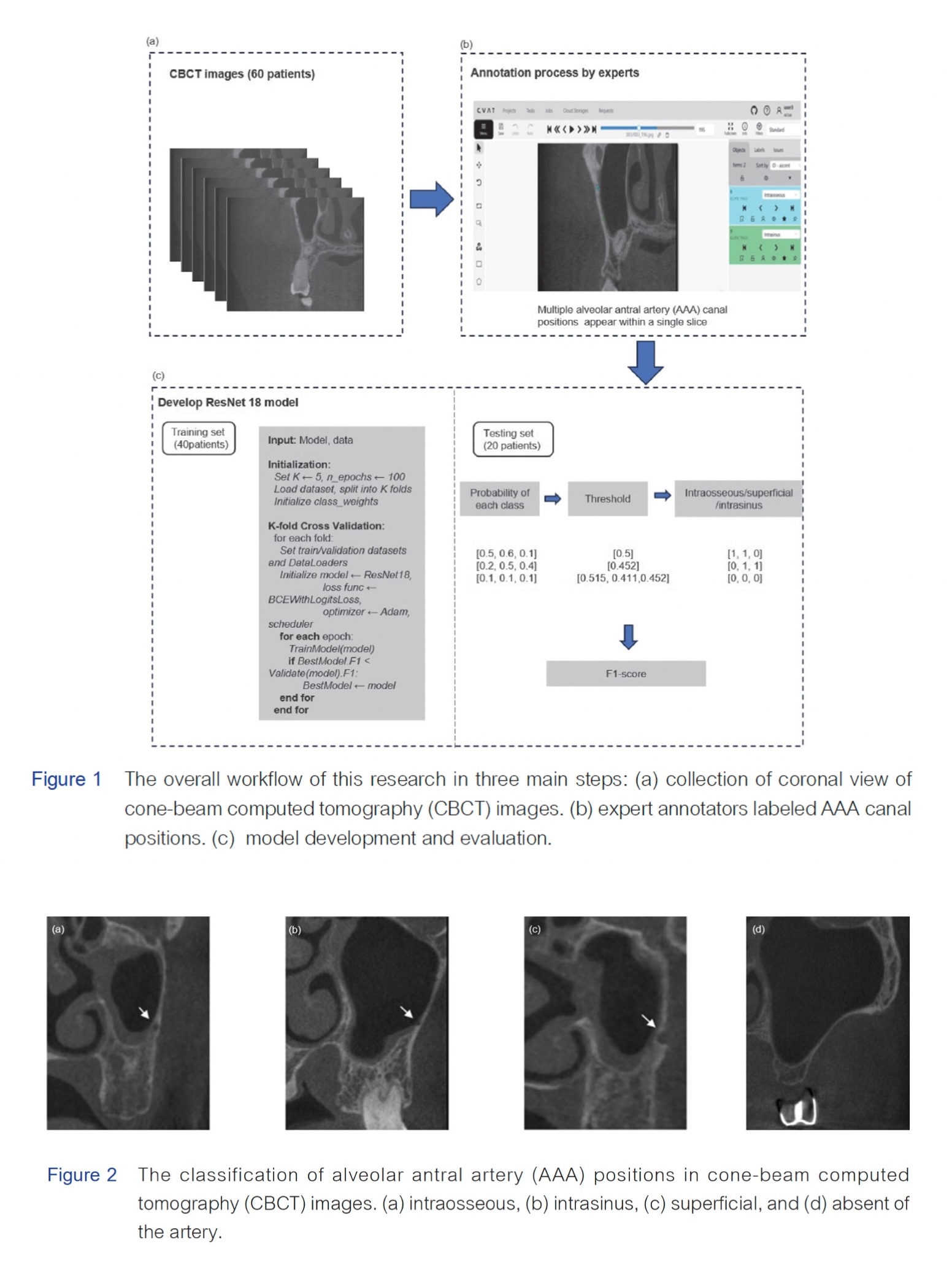
Objectives: The alveolar antral artery (AAA) supplies the posterior maxillary region. Detecting this artery using radiographs is essential before oral and maxillofacial surgeries to prevent bleeding complications. However, manual radiograph interpretation is time-consuming and requires expert experience. This study aims to develop and evaluate a ResNet-18 deep learning model that applies varying thresholds for multi-label classification of the AAA canal positions categorized as intraosseous, superficial, or intrasinus in coronal cone-beam computed tomography (CBCT) images.
Materials and Methods: Coronal CBCT images of 60 patients were selected and categorized into training data and testing data. Annotation of the AAA canal positions was done by experts via the Computer Vision Annotation Tool. Image transformation and augmentation techniques were applied to optimize a model training. A ResNet-18 model was trained with five-fold cross-validation. The best-performing model for each fold was determined based on the F1- score of the validation and tested on the test set.
Results: The model achieved a micro F1-score of 0.8206 when applying class-specific thresholds of 0.411, 0.452, and 0.515 for the superficial, intrasinus, and intraosseous positions, respectively. A single threshold of 0.452 and 0.500 marginally decreased the micro F1-scores to 0.8193 and 0.8157, respectively. The class-specific thresholds strategy yielded the highest per-class F1-scores, with values of 0.8153 for intraosseous, 0.6470 for intrasinus, and 0.0164 for the superficial class.
Conclusions: The implementation of deep learning for the AAA canal classification aids dentists and oral surgeons in preoperative planning, minimizing iatrogenic injury of the artery during surgery. Additionally, knowing the artery’s position beforehand enhances perioperative and postoperative management, allowing for better handling of complications in the event of vascular injury, potentially improving surgical outcomes and reducing procedural complications, benefiting both clinicians and patients.
Article Details

This work is licensed under a Creative Commons Attribution-NonCommercial-NoDerivatives 4.0 International License.
References
Larsen PE, Kennedy KS. Managing the posterior maxilla with implants using bone grafting to enhance implant sites. Oral Maxillofac Surg Clin North Am. 2019 May;31(2):299-308. doi:10.1016/j.coms.2019.01.002.
Molina A, Sanz-Sánchez I, Sanz-Martín I, Ortiz-Vigón A, Sanz M. Complications in sinus lifting procedures: Classification and management. Periodontol 2000. 2022 Feb;88(1):103-115. doi:10.1111/prd.12414.
Rosano G, Taschieri S, Gaudy JF, Weinstein T, Del Fabbro M. Maxillary sinus vascular anatomy and its relation to sinus lift surgery. Clin Oral Implants Res. 2011 Jul;22(7):711-715. doi:10.1111/j.1600-0501.2010.02045.x.
Danesh-Sani SA, Movahed A, ElChaar ES, Chong Chan K, Amintavakoli N. Radiographic evaluation of maxillary sinus lateral wall and posterior suerior alveolar artery anatomy: A cone-beam computed tomographic study Clin Implant Dent Relat Res. 2017 Feb;19(1):151-160. doi:10.1111/cid.12426.
Laovoravit V, Kretapirom K, Pornprasertsuk-Damrongsri S. Prevalence and morphometric analysis of the alveolar antral artery in a group of Thai population by cone beam computed tomography. Oral Radiol. 2021;37(3):452-462. doi:10.1007/s11282-020-00478-3.
Fahrettin Kalabalık HA. Evaluation of the alveolar antral artery position in the lateral sinus wall using cone-beam computed tomography. Ann Clin Anal Med 2020;11(4):330-334. doi: 10.4328/ACAM.20084
Zijderveld SA, van den Bergh JP, Schulten EA, ten Bruggenkate CM. Anatomical and surgical findings and complications in 100 consecutive maxillary sinus floor elevation procedures. J Oral Maxillofac Surg. 2008 Jul;66(7):1426-1438. doi:10.1016/j.joms.2008.01.027
Jensen SS, Eriksen J, Schiodt M. Severe bleeding after sinus floor elevation using the transcrestal technique: a case report. Eur J Oral Implantol. 2012 Autumn;5(3):287-291.
Yang DH, Lee NV. A simple method of managing the alveolar antral artery during sinus lift surgery. Int J Otolaryngol Head Neck Surg. 2021;10:131-146. doi: 10.4236/ijohns.2021.103014.
Varela-Centelles P, Loira M, González-Mosquera A, Romero-Mendez A, Seoane J, García-Pola MJ, et al. Study of factors influencing preoperative detection of alveolar antral artery by CBCT in sinus floor elevation. Sci Rep. 2020 Jul 2;10(1):10820. doi:10.1038/s41598-020-67644-9.
Testori T, Tavelli L, Scaini R, Saibene AM, Felisati G, Barootchi S, et al. How to avoid intraoperative and postoperative complications in maxillary sinus elevation. Periodontol 2000. 2023 Jun;92(1):299-328. doi:10.1111/prd.12480.
Pimkhaokham A, Aung CMS, Panmekiat S. The study of the alveolar antral artery canal in using cone beam computed tomography. M Dent J. 2016;37(1):63-69.
Ilgüy D, Ilgüy M, Dolekoglu S, Fisekcioglu E. Evaluation of the posterior superior alveolar artery and the maxillary sinus with CBCT. Braz Oral Res. 2013 Sep-Oct;27(5):431-437. doi:10.1590/s1806-83242013000500007.
Ketabi AR, Hassfeld S, Lauer HC, Piwowarczyk A. Comparative diagnosis of the alveolar antral artery canal in the lateral maxillary sinus wall in corresponding panoramic radiography and cone-beam computed tomography. Int J Implant Dent. 2023 Sep 19;9(1):30. doi:10.1186/s40729-023-00497-9.
Kqiku L, Biblekaj R, Weiglein AH, Kqiku X, Städtler P. Arterial blood architecture of the maxillary sinus in dentate specimens. Croat Med J. 2013 Apr;54(2):180-184. doi:10.3325/cmj.2013.54.180.
Solar P, Geyerhofer U, Traxler H, Windisch A, Ulm C, Watzek G. Blood supply to the maxillary sinus relevant to sinus floor elevation procedures. Clin Oral Implants Res. 1999 Feb;10(1):34-44. doi:10.1034/j.1600-0501.1999.100105.x.
Ossowska A, Kusiak A, Świetlik D. Artificial Intelligence in dentistry-narrative review. Int J Environ Res Public Health. 2022 Mar 15;19(6):3449. doi: 10.3390/ijerph19063449.
Anaya-Isaza A, Mera-Jiménez L, Zequera-Diaz M. An overview of deep learning in medical imaging. Inform Med Unlocked. 2021;26:100723. doi:10.1016/j.imu.2021.100723.
Xu W, Fu YL, Zhu D. ResNet and its application to medical image processing: Research progress and challenges. Comput Methods Programs Biomed. 2023 Oct;240:107660. doi:10.1016/j.cmpb.2023.107660.
He K, Zhang X, Ren S, Sun J. Deep residual learning for image recognition. 2016 IEEE Conference on Computer Vision and Pattern Recognition (CVPR). Las Vegas, NV, USA, 2016, pp.770-778, doi: 10.1109/CVPR.2016.90.
Yang Y, Zhang L, Du M, Bo J, Liu H, Ren L, et al. A comparative analysis of eleven neural networks architectures for small datasets of lung images of COVID-19 patients toward improved clinical decisions. Comput Biol Med. 2021 Dec;139:104887. doi:10.1016/j.compbiomed.2021.104887.
Huang G, Liu Z, Maaten LVD, Weinberger KQ, editors. Densely connected convolutional networks. 2017 IEEE Conference on Computer Vision and Pattern Recognition (CVPR). Honolulu, USA, 2017, pp.4700-4708. doi.org/10.1109/CVPR.2017.243
Wu Z, Zhuo R, Liu X, Wu B, Wang J. Enhancing surgical decision-making in NEC with ResNet18: a deep learning approach to predict the need for surgery through x-ray image analysis. Front Pediatr. 2024 Jun 4;12:1405780. doi:10.3389/fped.2024.1405780.
Jiang T, Lu Z, Hu X, Zeng L, Ma X, Huang J, et al. Deep learning multi-label tongue image analysis and its application in a population undergoing routine medical checkup. Evid Based Complement Alternat Med. 2022 Sep 29;2022:3384209. doi:10.1155/2022/3384209.
Kwak GH, Kwak EJ, Song JM, Park HR, Jung YH, Cho BH, et al. Automatic mandibular canal detection using a deep convolutional neural network. Sci Rep. 2020 Mar 31;10(1):5711. doi:10.1038/s41598-020-62586-8.
Jaskari J, Sahlsten J, Järnstedt J, Mehtonen H, Karhu K, Sundqvist O, et al. Deep learning method for mandibular canal segmentation in dental cone beam computed tomography volumes. Sci Rep. 2020 3 Apr;10(1):5842. doi:10.1038/s41598-020-62321-3.
Choi H, Jeon KJ, Kim YH, Ha E-G, Lee C, Han S-S. Deep learning-based fully automatic segmentation of the maxillary sinus on cone-beam computed tomographic images. Sci Rep. 2022 Aug 17;12(1):14009. doi:10.1038/s41598-022-18436-w.
Rahpeyma A, Khajehahmadi S. Alveolar antral artery: review of surgical techniques Iivolving this anatomic structure. Iran J Otorhinolaryngol. 2014;26(75):73-78.
Maridati P, Stoffella E, Speroni S, Cicciù M, Maiorana C. Alveolar antral artery isolation during sinus lift procedure with the double window technique. Open Dent J. 2014 May30;8:95-103. doi:10.2174/1874210601408010095.
Park JA, Kim D, Yang S, Kang JH, Kim JE, Huh KH, et al. Automatic detection of posterior superior alveolar artery in dental cone-beam CT images using a deeply supervised multi-scale 3D network. Dentomaxillofac Radiol. 2024 Jan 11;53(1):22-31. doi:10.1093/dmfr/twad002.
Putra RH, Doi C, Yoda N, Astuti ER, Sasaki K. Current applications and development of artificial intelligence for digital dental radiography. Dentomaxillofac Radiol. 2022 Jan1;51(1):20210197. doi:10.1259/dmfr.20210197.
Schwendicke F, Samek W, Krois J. Artificial intelligence in dentistry: chances and challenges. J Dent Res. 2020 Jul;99(7):769-774. doi:10.1177/0022034520915714.
Staněk J, Machálková K, Staňková M, Zapletalová J, Kocurová T. Alveolar antral artery: cone beam computed tomography study and clinical context. PeerJ. 2023 Nov 30;11:e16439. doi:10.7717/peerj.16439.
Luque A, Carrasco A, Martín A, de las Heras A. The impact of class imbalance in classification performance metrics based on the binary confusion matrix. Pattern Recognit. 2019 Jul;91:216-231. doi:10.1016/j.patcog.2019.02.023.
Saito T, Rehmsmeier M. The precision-recall plot is more informative than the ROC plot when evaluating binary classifiers on imbalanced datasets. PLoS One. 2015 Mar4;10(3):e0118432. doi:10.1371/journal.pone.0118432.
Benjaphalakron N, Jansisyanont P, Chuenchompoonut C, Kiattavorncharoen S. Evaluation of the posterior superior alveolar artery and related factors using cone beam computed tomography images. JDAT 2021;71:35-43.
Ragab MG, Abdulkadir SJ, Muneer A, Alqushaibi A, Sumiea EH, Qureshi R, et al. A comprehensive systematic review of YOLO for medical object detection (2018 to 2023).IEEE Access 2024 Apr.;12:57815 - 57836. doi: 10.1109/ACCESS.2024.3386826.
